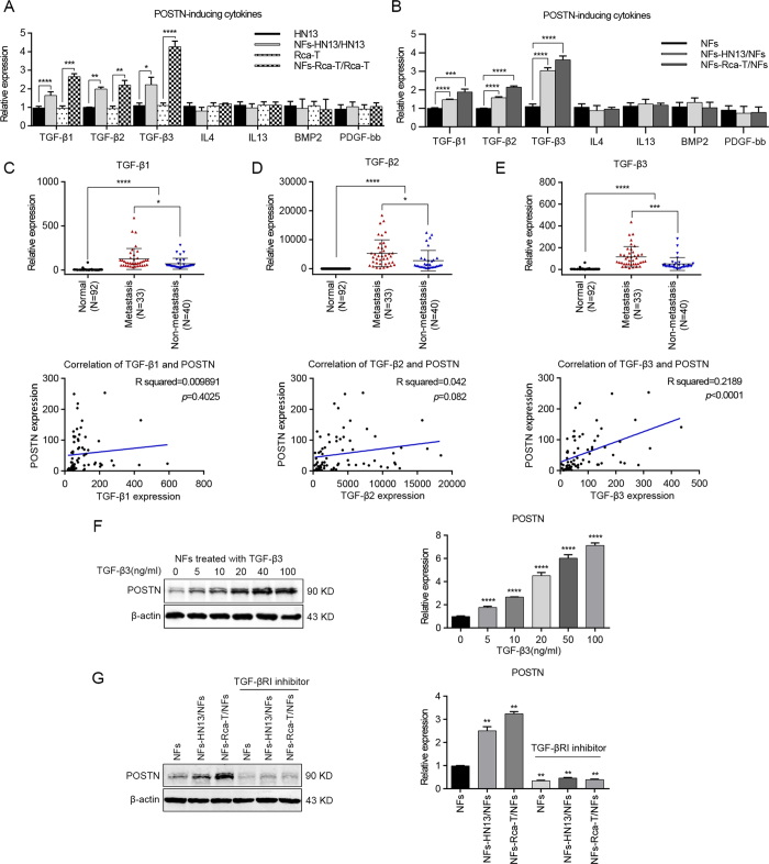
Figure 6. TGF-β3 induced POSTN expression in NFs after co-culture with HNC cells.
(A,B) The mRNA levels of TGF-β1, TGF-β2, TGF-β3, IL-4, IL-13, BMP2 and PDGF-bb in HNC cells (A) and NFs (B) were detected after the co-culture of NFs and HNC cells by real-time PCR. (C–E) TGF-β1, TGF-β2 and TGF-β3 mRNA levels were determined using real-time PCR in 73 cases of HNC tissues (involving 33 metastatic cases) and in 92 cases of normal oral epithelial tissues. A significant positive correlation was observed between the TGF-β3 and POSTN expression levels in the HNC tissues (n = 73). (F) TGF-β3 induced POSTN expression in NFs in a dose-dependent manner. Protein expression by western blot analysis and mRNA expression by real-time PCR were measured at the indicated concentrations. (G) Blocking of TGF-β3 with the inhibition of type I TGF-β receptor (SB431542, 10 μM) abrogated the induced-POSTN expression in NFs after the co-culture of NFs and HNC cells. POSTN protein and mRNA levels were determined in NFs using western blotting and real-time PCR. (*p < 0.05; ** p < 0.01; ***p < 0.001; ****p < 0.0001).

