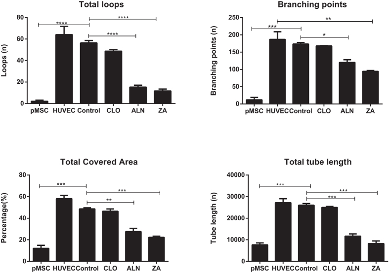
Figure 8. Quantitative analysis of the morphological tube formation parameters; total covered area, total loop length, loop numbers and the number of branching points (Wimasis WimTube, Wimasis GmbH Munich, Germany) were all significantly reduced in the pMSC derived endothelial cells in the presence of BPs compared to both stimulated pMSCs and HUVECs.
Inhibition was potency dependent with the most significant inhibition seen in the ALN and ZA treated cells. (Significant differences, compared to EGMV are indicated as *p < 0.05, **p < 0.01, ***p < 0.001 and ****p < 0.0001).

