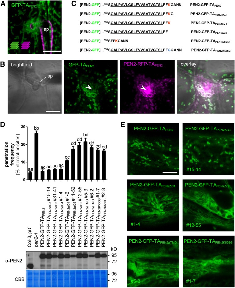
Figure 2.
The C-Terminal Extension of PEN2 Is Important for Subcellular Localization and Functionality.
(A) Maximum z-projection of CLSM images from a transgenic leaf epidermal cell expressing GFP-TAPEN2 reveal localization of the truncated protein to the periphery of subcellular membrane compartments 24 hpi with Bgh. Fungal structures were stained with FM4-64 (z-stack 22 µm). ap, appressorium. Bar = 10 µm.
(B) Single CLSM image of transgenic leaf epidermal cells coexpressing GFP-TAPEN2 and full-length PEN2-RFP-TAPEN2 shows colocalization of both proteins in the same membrane compartment, but GFP-TAPEN2 does not contribute to PEN2-RFP-TAPEN2 hyperfluorescence underneath the fungal appressorium 22 hpi with Bgh. Arrowheads point to PEN2-RFP-TAPEN2 hyperfluorescence at the site of attempted fungal invasion. ap, appressorium. Bar = 10 µm.
(C) Protein sequence of the C-terminal extension of different PEN2 TA deletion constructs. The predicted transmembrane domain is underlined. Positively charged amino acid lysine is marked in red. Number indicates protein position in the native PEN2 protein.
(D) and (E) pen2-1 mutant complementation analyses and CLSM of PEN2-GFP TA deletion constructs indicate full complementation capacity only occurs for proteins that associate with membrane compartments.
(D) Upper part: Frequency of invasive growth at Bgh interaction sites 72 hpi on Col-3, gl1, pen2-1, and plants expressing full-length PEN2-GFP-TAPEN2 or different deletion proteins. Different letters indicate significantly different classes (99% confidence intervals) determined by one-way ANOVA with Tukey’s post test. Depicted results represent two biological replicates with 100 interaction sites analyzed on three different leaves each. Error bars indicate se of the mean. Lower part: Corresponding immunoblot analyses of all plant lines using 30 µg protein and the PEN2-specific antibody show different levels of protein expression. CBB, Coomassie Brilliant Blue staining. Experiments were repeated three times with similar results.
(E) Single CLSM images of transgenic plants show subcellular localization of PEN2-GFP-TAPEN2 and different PEN2-GFP TA deletion constructs. The deletion proteins PEN2-GFP-TAPEN2∆C5, PEN2-GFP-TAPEN2∆TMD, and PEN2-GFP-TAPEN2K556G show diffuse distribution in the cytoplasm and, possibly, reticulate ER localization. Bar = 10 µm.

