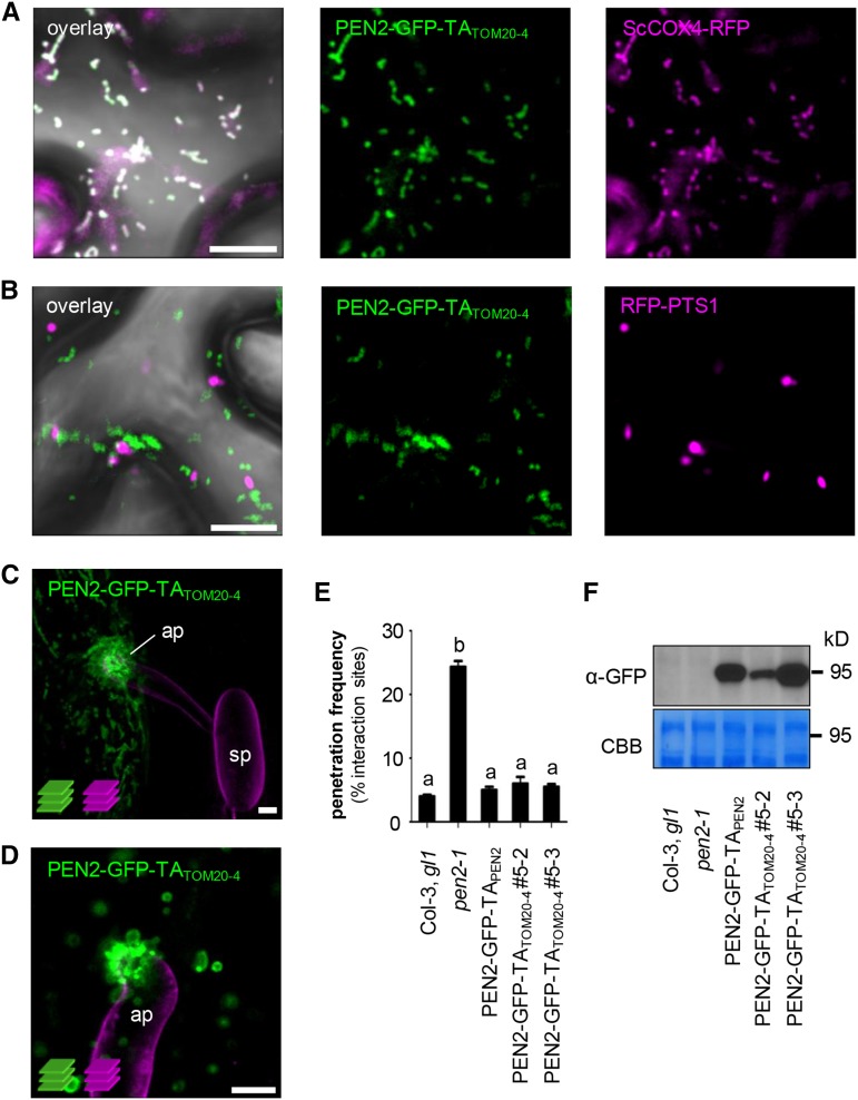
Figure 4.
The C-Terminal TA of TOM20-4 Can Functionally Replace PEN2’s TA for Proper Subcellular Localization and Functionality.
(A) and (B) CLSM images of N. benthamiana leaf epidermal cells, transiently coexpressing PEN2-GFP-TATOM20-4 and either RFP-tagged mitochondrial marker (A) or RFP-labeled peroxisomes (B), show that PEN2-GFP-TATOM20-4 is exclusively associated with mitochondria and not peroxisomes 3 d after Agrobacterium tumefaciens infiltration.
(C) and (D) Two examples of z-projections of CLSM images from transgenic leaf epidermal cells expressing PEN2-GFP-TATOM20-4 exhibit the same subcellular behavior and aggregate formation as PEN2-GFP-TAPEN2 20 hpi with Bgh. Fungal structures were stained with FM4-64 (z-stack 22 and 9 µm). ap, appressorium; sp, spore. Bars = 10 µm.
(E) Complementation analyses of pen2-1 mutant by PEN2-GFP-TATOM20-4 indicate full complementation capacity of the chimeric construct. Results were scored 72 hpi with Bgh. Different letters indicate significantly different classes (99% confidence intervals) determined by one-way ANOVA with Tukey’s post test. Depicted results represent two biological replicates with 100 interaction sites analyzed on three different leaves, each. Error bars indicate se of the mean.
(F) Corresponding immunoblot analyses of all plant lines using 30 µg protein extract and a GFP-specific antibody show different levels of protein expression. Experiments have been repeated three times with similar results. CBB, Coomassie Brilliant Blue staining.

