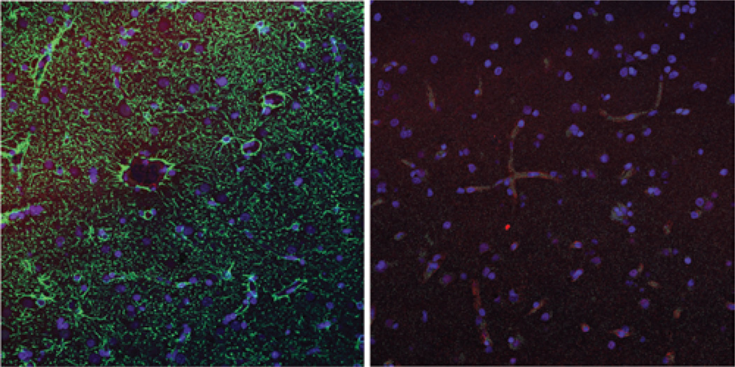
Fig. 4. Nonenhancing tissue.
Left: Nonenhancing region of glioblastoma multiforme (Case 5). The green immunofluorescence represents GFAP staining, indicating glial processes which wrap tightly around the CD31-positive (red) vasculature. Right: Sample from nonenhancing brain tissue resected together with a metastasis (Case 9), with red immunofluorescence showing AQ4 staining tightly around the green CD31-positive vessels.

