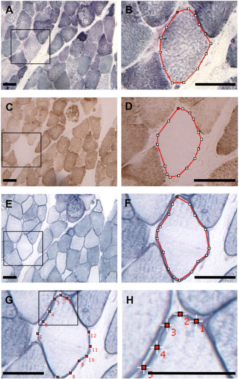
Figure 1. Histochemical quantification in skeletal muscle.
Muscle section from a patient with SDH (A,B), COX (C,D) and NADPHd (E–H) histochemistry. We show an example of a COX− fibre with reduced sarcoplasmic NOS and increased sarcolemmal NOS activities. SDH (B), COX (D) and sarcoplasmic NOS (E,F) activities were measured by circumscribing (red line) the sarcoplasm, with an interactive cursor, excluding the sarcolemmal membrane in the case of sarcoplasmic NOS activity. Sarcolemmal NOS activity (G,H) was obtained by placing fixed size squares at 12 different sites on the sarcolemmal membrane. Care was taken to avoid areas with superimposed membranes. The mean optical density (O.D) measured inside the squares and circumscribed areas was considered as an estimate of the histochemical activity. Panels (B,D,F,H) are amplified images delimitated by the black square frame on (A,C,E,G), respectively. SDH: succinate dehydrogenase, COX: cytochrome-c-oxidase, NADPHd: NADPH diaphorase, COX− = COX negative. Scale bar = 50 μm.

