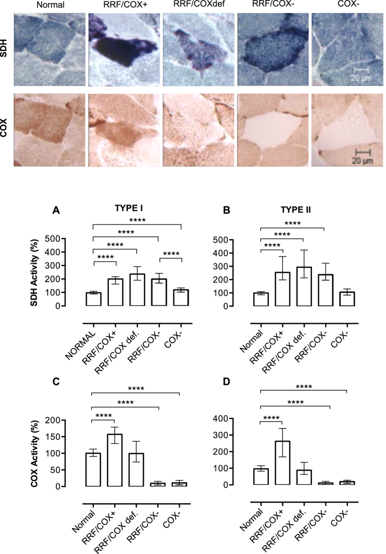
Figure 2. Types of abnormalities in muscle fibres after SDH and COX histochemistry.
The panel on top shows examples of the different types of muscle fibres included in this study on SDH and COX histochemistry. From the left to the right, we demonstrate an example of normal muscle and each type of abnormal fibres: fibre with mitochondrial proliferation and preserved COX activity (RRF/COX+), fibre with mitochondrial proliferation and impaired COX activity (RRF/COXdef), fibre with mitochondrial proliferation and low COX activity (RRF/COX−), fibre with low COX activity and without mitochondrial proliferation (COX−). SDH quantification shows that all fibres with mitochondrial proliferation (RRF/COX+, RRF/COXdef, RRF/COX−) have higher values when compared to normal fibres (A,B). Type I COX− fibres have also increased SDH staining, but is significantly different when compared to RRF/COX− fibres showing that it is a distinct group. COX quantification (C,D) demonstrates that RRF/COX+ fibres have increased COX activity while RRF/COX− and COX− fibres had lower activities. The total numbers of myofibres analysed in each group are shown in Table 2. Data were analysed by Kruskal-Wallis test followed by Dunn’s post hoc test. ****P ≤ 0.0001. Bars are showing median and interquartile range.

