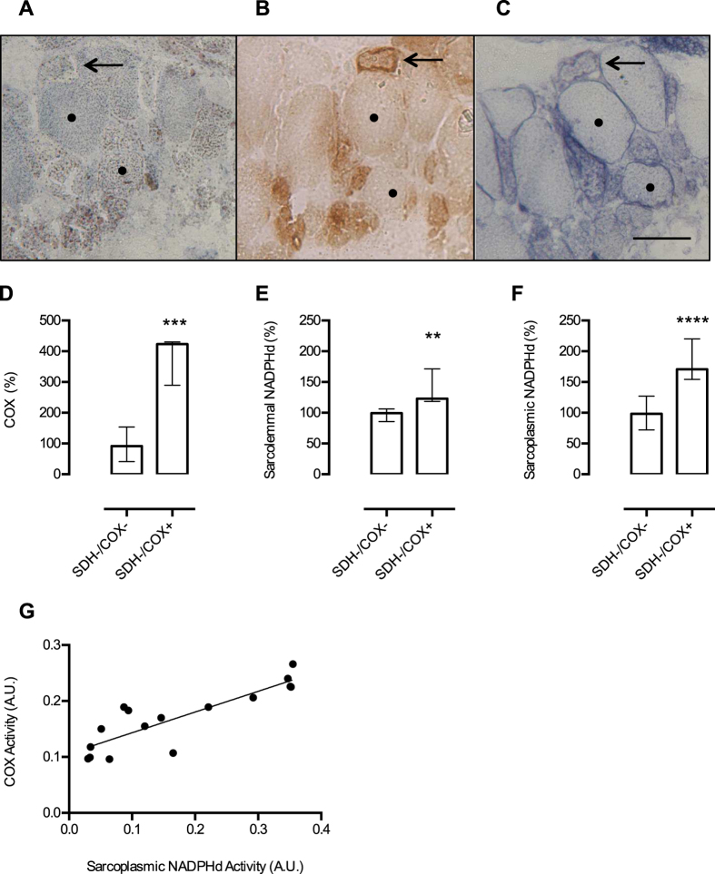
Figure 5. NADPHd activity in muscle fibres and Complex II deficiency.
Muscle biopsy from a patient with a complex pattern of mitochondrial enzyme deficiency (complex I, II and IV deficiencies) shows a diffuse lack of SDH staining (A). COX histochemistry (B) shows fibres with low (•) and increased (arrow) COX activities, showing that mitochondrial content was increased (RRF). NADPHd (C) shows that despite SDH deficiency, the same pattern observed in other patients is present: low sarcoplasmic NOS activity in COX− fibre (•) and increased sarcolemmal and sarcoplasmic NOS activities in RRF/COX+ fibre (arrow). Based on COX activity, we separated the fibres in two groups: SDH-/COX− (n = 10) and SDH-/COX+ (n = 8). After quantification of COX staining, we show that the group of SDH-/COX+ fibres has significantly increased COX activity (P = 0.0005; D). NADPHd activities were increased in both sarcolemmal (P = 0.0014; E) and sarcoplasm (P < 0.0001; F) in SDH-/COX+ muscle fibres. There is a good correlation between COX and sarcoplasmic NADPHd activities (n = 16; r = 0.86; G). A.U. = arbitrary units. Data presented in D, E, and F were analyzed by Mann-Whitney test, ** P ≤ 0.01, ***P ≤ 0.001, ****P ≤ 0.0001. Scale bar = 50 μm. Bars are showing median and interquartile range.

