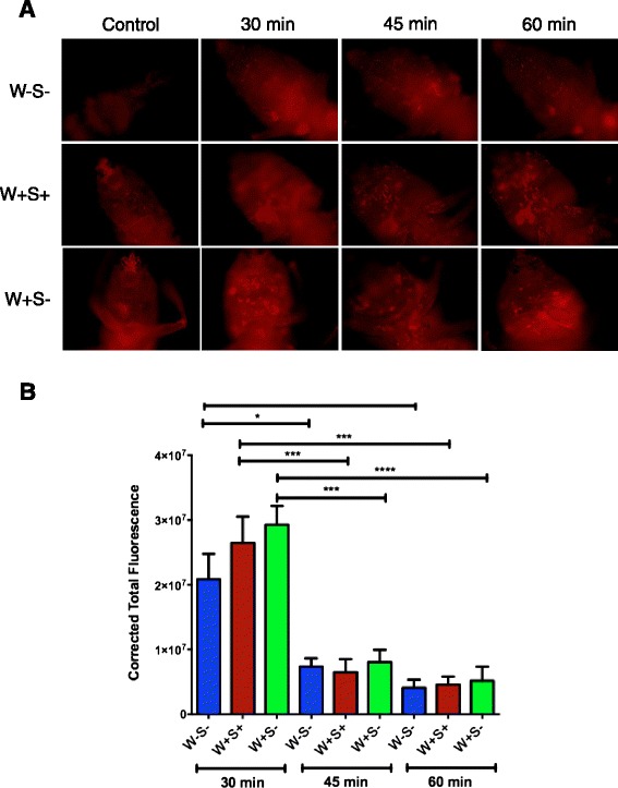
Fig. 6.

Phagocytosis in D. melanogaster flies carrying or lacking endosymbiotic bacteria. a Representative images of phagocytosis in D. melanogaster adult flies without Wolbachia and Spiroplasma (W-S-), with both endosymbionts (W + S+), and with Wolbachia only (W + S-) at 30, 45 and 60 min after injection of lipophilized pHrodo-labeled E. coli particles. Control treatments involved injections with DEPC-treated water. Images were taken using fluorescence microscopy and 10x magnification. b Corrected total fluorescence in the three D. melanogaster flies at 30, 45 and 60 min following injection of pHrodo-labeled E. coli. Images were processed in ImageJ and corrected total fluorescence was estimated by measuring relative amounts of fluorescence, which included estimations of the resulting area, mean fluorescence of background and integrated density. The experiment was repeated three times with 6-12 flies for each treatment. ****P < 0.0001; ***P < 0.001; **P < 0.01; *P < 0.05 (one way analysis of variance with a Tukey post hoc test, GraphPad Prism5 software)
