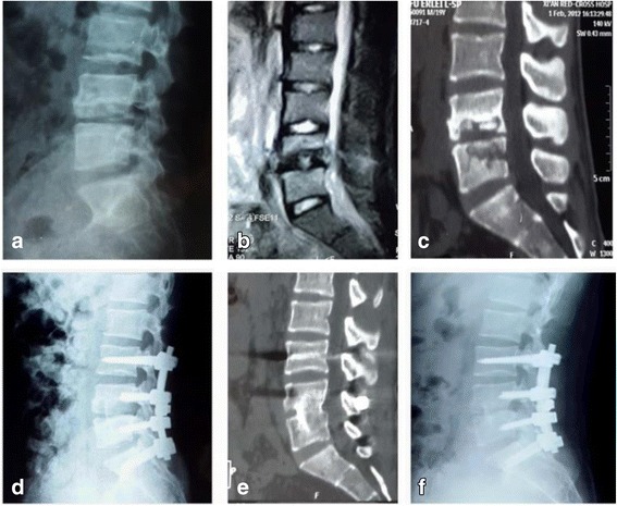
Fig. 1.

Images from a 21-year-old man with lumbar spinal tuberculosis who underwent one-stage posterior debridement, bone grafting, and instrumentation. a–c Preoperative X-ray, MR, and CT images showing the destruction of L4 and L5 and paravertebral abscess formation. d Postoperative lateral X-ray showing fixation of L3–S1. e, f At the 24-month follow-up examination, fixation and interbody vertebral fusion were satisfactory, with no sign of tuberculosis recurrence
