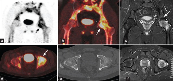
Figure 3.
A 39-year-old male patient with left sided hip pain for 1-year: (a) Coronal positron emission tomography, (b) Fused coronal positron emission tomography/computed tomography, (d) Axial fused positron emission tomography/computed tomography images show diffusely increased tracer uptake (arrow) in the head and neck of the left femur, (e) Computed tomography showed no morphological changes (c and f) coronal and axial magnetic resonance imaging images show diffuse bone marrow edema extending up to the neck and decreased signal intensity from the 11–2 O’clock position (arrow) in the left femoral head. A diagnosis of transient osteoporosis of the hip was made which was proved on clinical follow-up

