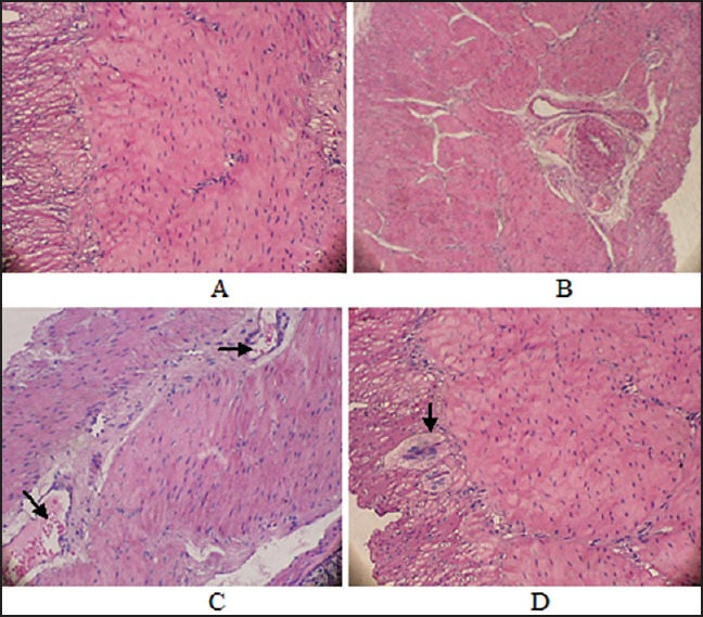
Figure 5.

Histology of pyloric submucosas (H&E, ×40). (a) Control group (b and c) Infected groups (b) Dilated congested blood vesicles with cellular infiltration and edematous aria between muscles (fibrosis) (c) Cellular infiltration increased (d) Treated group showing nerve hyperplasia and a decrease in dilation of the blood vesicles and disappearance of cellular infiltration
