Fig. 3.

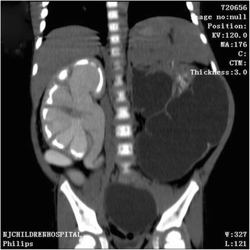
Contrast-enhanced computed tomography scan shows renal dysplasia with giant ureter on the left side and right hydronephrosis with the whole right ureter dilatation

Contrast-enhanced computed tomography scan shows renal dysplasia with giant ureter on the left side and right hydronephrosis with the whole right ureter dilatation