
Fig. 1.
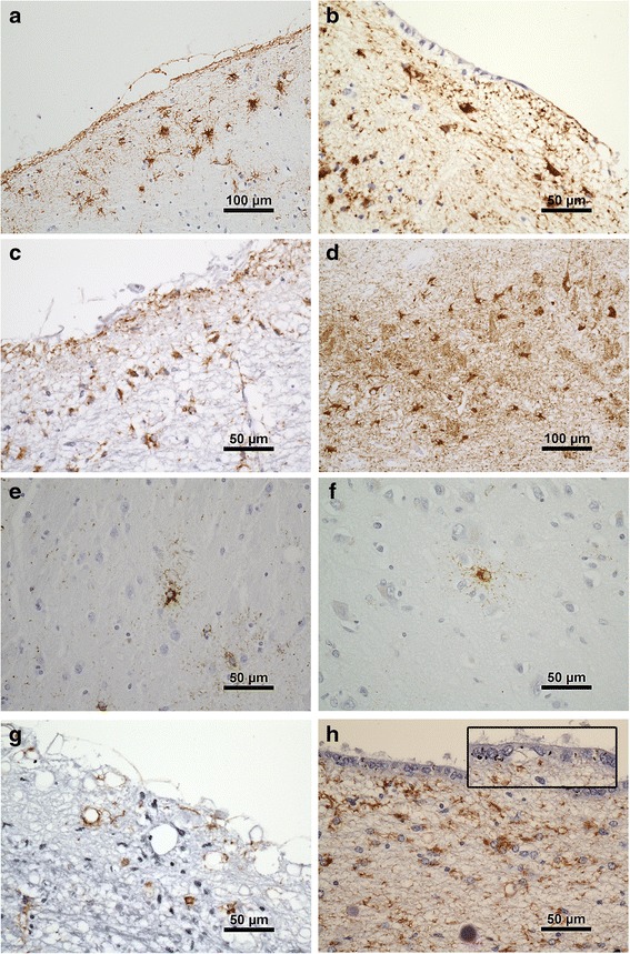
Examples of tau pathologies in the cohort. a. Subpial TSA temporal cortex. Note layer of subpial tau + neurites (arrow). b. Subependymal TSA hippocampal region. c. Midbrain subpial TSA and neurites. d. TSA in CA2 region of hippocampus. e. Astrocyte with finely tau immunoreactive processes in striatum and, f. in temporal cortex. g. Tau + neurites pons, without TSA. h. Tau positive neurites in subependymal and ependymal (arrow and inset) location associated with TSA, hippocampal region