
Fig. 1.
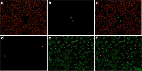
Immunofluorescence staining of Iba-1 (a, d red) in microglia and GFAP (b, e green) and merged images (c, f) in astrocytes in the microglial (a–c) and astrocytic cultures (d–f). The results suggested that more than 99 % of cells prepared from human brains are microglia and astrocytes, respectively. The calibration bar in F: 50 μm