
Fig. 1. The electron transport chain in mycobacteria and synthesis of menaquinone.
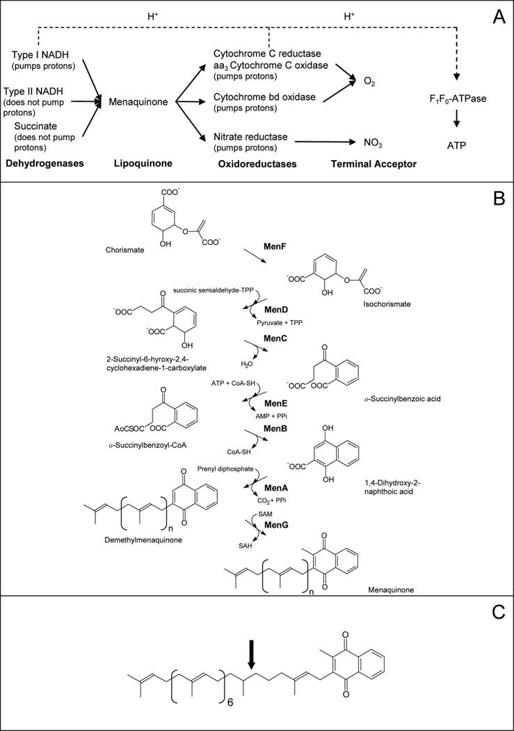
Panel A: Architecture of selected aerobic respiratory pathways in mycobacteria. Panel B: Biosynthetic pathway for menaquinone in E. coli. Enzyme names are indicated in bold. C: Structure of the major menaquinone species found in mycobacteria, MK-9 (II-H2) (Minnikin, 1982); calculated monoisotopic mass = 786.63148. Menaquinones are identified by the length and chemical structure of prenyl chain, the predominant form of menaquinone in mycobacteria has 9 isoprene units with the second one being saturated (indicated by the arrow). Hence this menaquinone is identified as MK-9 (II-H2). The fully unsaturated version is known as MK-9 and has a calculated monoisotopic mass of 784.61583