
Fig. 6. Comparison of the levels of Δ9-desaturase activity in M. bovis BCG wild type and M. bovis BCG/pVV16desA3.
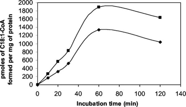
A final volume of 1 ml of reaction mixture contained crude cell lysate of M. bovis BCG wild type (diamonds) or BCG/pVV16desA3 (squares), 20 μl of solution containing 50% Me2SO and 1% Tween 20, 1 μmol of NADPH in 0.1 mM potassium phosphate buffer, pH 7.2, and 25 nmol of [1-14C]18: 0-CoA as substrate. Duplicate assays were performed. After the indicated incubation times, the reactions were stopped with 1 ml of 15% tetrabutylammonium hydroxide. FAMEs were extracted, methylated, and analyzed for[1-14C]C18:1 by argentation TLC and the Bioscan System.