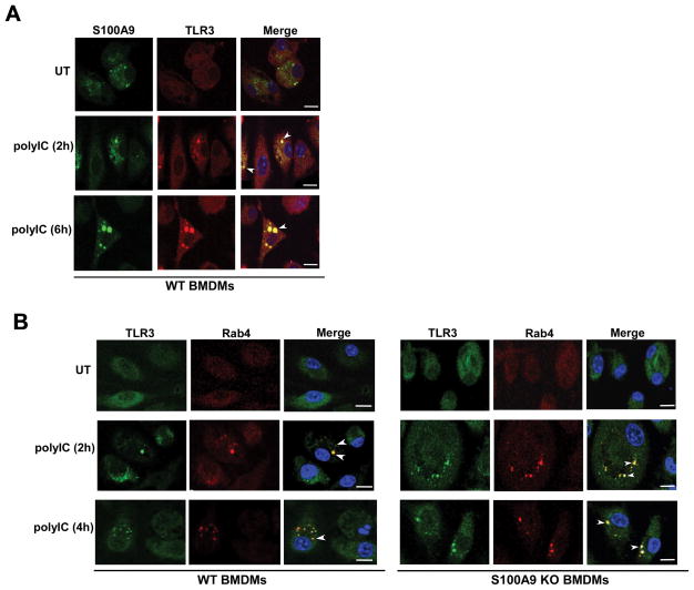
FIGURE 5. S100A9 co-localizes with TLR3 upon polyIC stimulation and S100A9 is not required for TLR3 trafficking to early endosome.
(A) Co-immunofluorescence (co-IFA) analysis was performed by treating primary bone marrow derived macrophages (BMDM) isolated from wild-type (WT) mice with polyIC. Following treatment, cells were fixed and labeled with S100A9 (FITC- green) and TLR3 (Texas red - red) antibodies. The images of polyIC treated cells represent 70% of cells (i.e. 70 cells out of 100 cells) with S100A9 and TLR3 co-localization. (B) BMDM isolated from WT and S100A9 knockout (KO) mice were treated with polyIC for co-IFA analysis. Following polyIC treatment, cells were fixed and labeled with antibodies specific for TLR3 (FITC- green) and early endosome marker Rab4 (Texas red - red). The images of polyIC treated WT and S100A9 KO cells represent 68% and 72% of cells (i.e. 68 cells or 72 cells out of 100 cells) with Rab4 and TLR3 co-localization, respectively. Merged images in (A) and (B) are represented as yellow. The images are representative of thirty viewing fields from two independent experiments with similar results. White arrow heads in merged images represents co-localization. The white bar represents 10 μm. UT; untreated cells.

