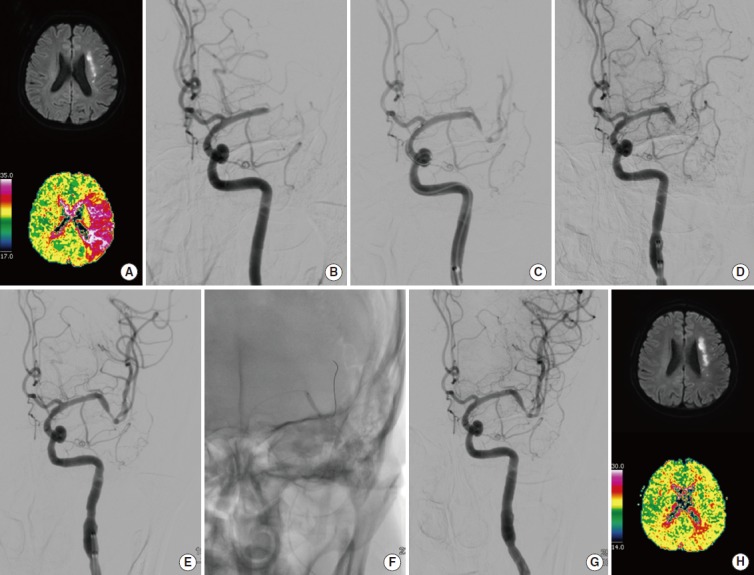
Figure 1.

A 56-year-old man (patient No. 3) presented with left hemispheric syndrome. Baseline magnetic resonance imaging (MRI) shows an acute left internal borderzone infarction on diffusion-weighted imaging, and a large perfusion defect on time-to-peak of perfusion-weighted imaging (A). Initial cerebral angiography shows occlusion of the distal M1 segment of the left middle cerebral artery (B). Temporary blood flow bypass is seen when the Solitaire FR stent is being deployed (C). Although some clots were removed at the occlusion site, occlusion is still seen at a more distal vessel (D). After 3 stent retrieval passes, partial recanalization was achieved, but flow still seems stagnated (E). After 2 angioplasties (F), complete reperfusion was fully achieved (G). Repeat MRI performed on the fifth day of admission shows similar infarct volume and complete recovery of the perfusion defect (H). The patient’s modified Rankin Scale score was 1 at 3 months and 0 at 1 year.
