
Fig. 1. Early lethality of perinatal cardiomyocyte-specific Parkin-deficient mice.
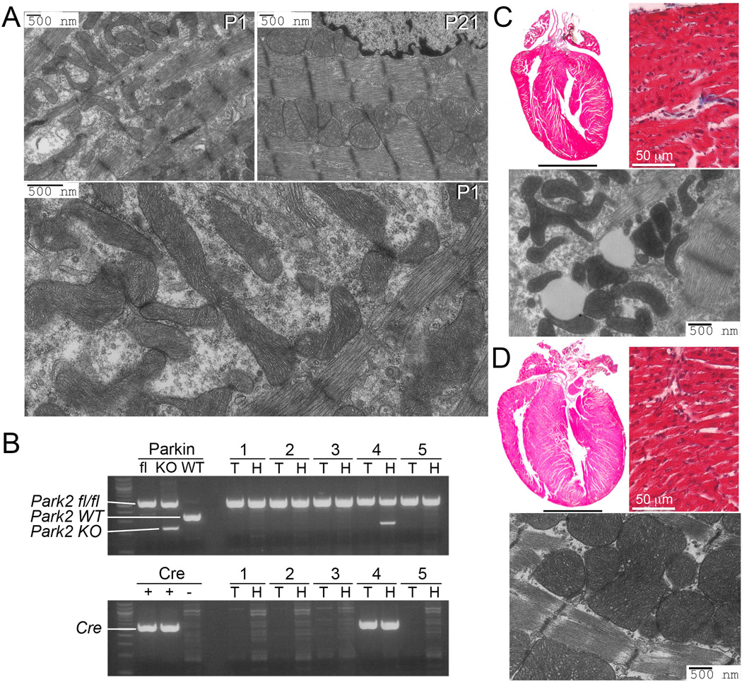
(A) Transmission electron microscopy (TEM) showing normal cardiomyocyte mitochondria on the first (P1) and 21st (P21) day of life. Enlargement shows structural details at P1. (B) PCR genotyping of floxed Park2 gene (top) and tamoxifen-inducible cardiac Cre (bottom) of surviving mice from a representative litter at P21; 3 mice died before weaning. KO indicates Cre recombined Park2 fl/fl allele. T is tail DNA; H is heart DNA. (C and D) Representative (of 3) hearts, histological sections, and TEMs from P21 cardiac Parkin deficient (top) and control (bottom) mice. Scale bars for hearts is 2 mm.