
Fig. 3. Perinatal cardiomyopathy evoked by non-phosphorylated Mfn2 AA.
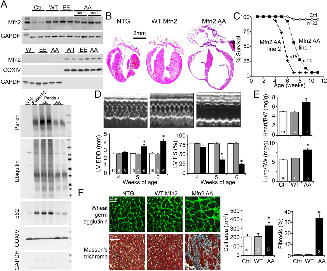
(A) Immunoblot analysis of Mfn2 expression (top) and mitochondrial Parkin localization (bottom) in transgenic mouse hearts. Upper panel - top pair is cardiac homogenate; bottom pair is mitochondrial-enriched 10,000g pellet (cytochrome oxidase IV; COX IV) and post-mitochondrial supernatant (GAPDH). Lower panel - Immunoblot analysis of mitochondrial-associated Parkin and downstream mitophagy events and their modulation by cardiac-expressed Mfn2 EE and Mfn2 AA. (B) Representative hearts of 6 week old mice. (C) Survival. (D) Serial echocardiographic data of 4-6 week old mice; white bars are Ctrl, grey is WT Mfn2, and black is Mfn2 AA. (E) Heart (top) and lung (bottom) weights of 6 week old mice indexed to body weight (BW). (F) Histological studies of cardiomyocyte cross sectional area (top) and myocardial fibrosis (bottom); quantitative data are on the right. *is p<0.05 vs WT Mfn2 and NTG control.