
Fig. 5. Fetal mitochondria persist in young adult Mfn2 AA mouse hearts.
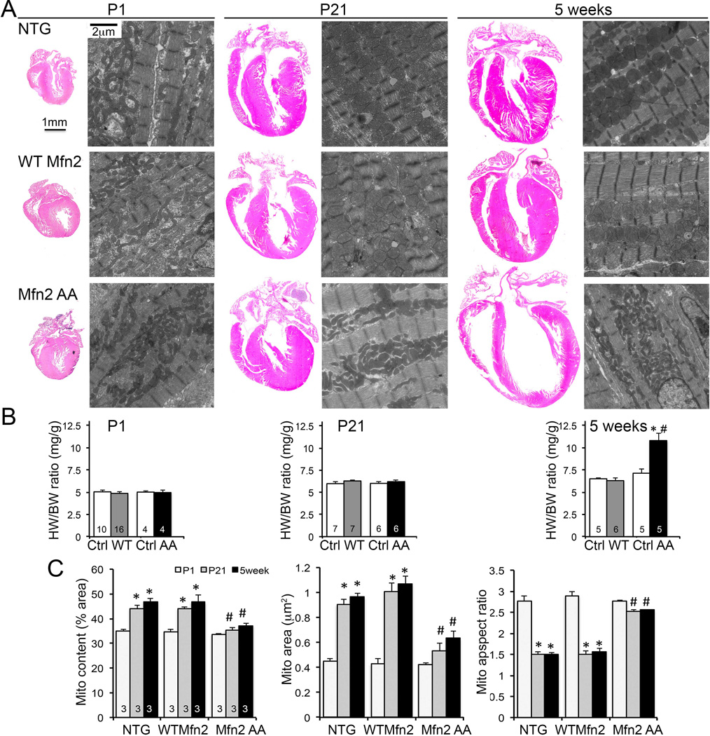
(A) Representative 4-chamber heart sections and transmission electron micrographs of cardiomyocyte mitochondria from P1, P21, and 5 week old mouse hearts. NTG controls are top row, WT Mfn2 middle row, Mfn2 AA bottom row. Quantitative data for heart weights are in (B) and for mitochondrial ultrastructure in (C); * = p<0.05 vs P1, # = p<0.05 vs WT Mfn2 at same stage.