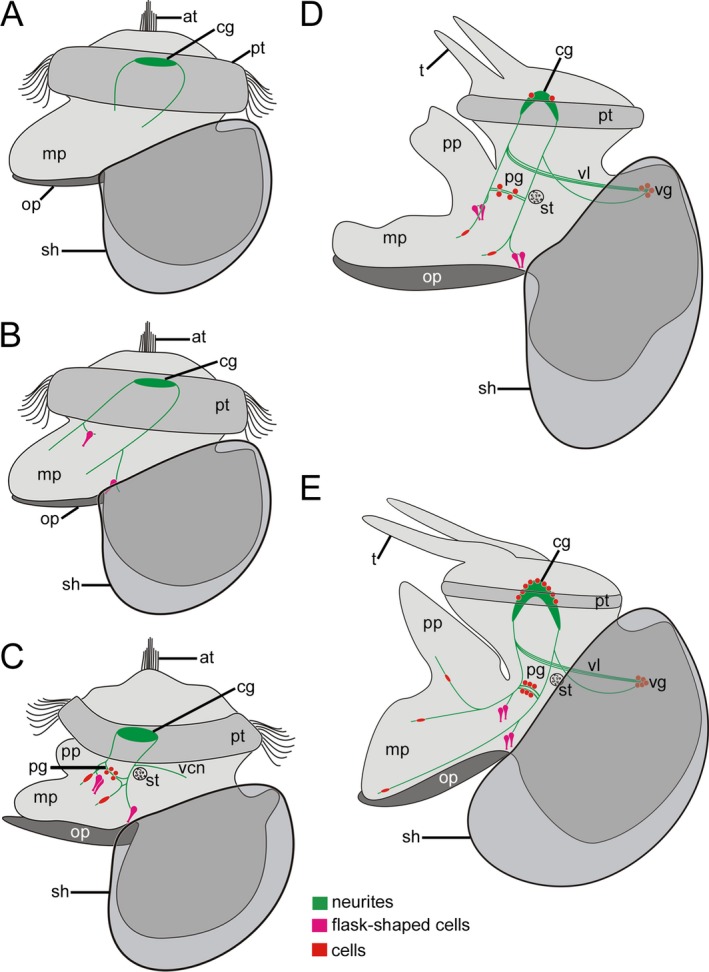
Figure 7.

Schematic representation of FMRFamide‐like immunoreactivity (lir) in Lottia cf. kogamogai. Anterior faces upwards in all images. (A‐E) lateral view, ventral to the left. Total size of specimens is approx. 120 μm in (A); 125 μm in (B); 140 μm in (C); 170 μm in (D); 180 μm in (E). Age of larvae is given in hours or days postfertilization (hpf, dpf). (A) neurites are the first labelled neuronal structures that appear in the future adult cerebral ganglia and cerebropedal connectives in the late veliger larva (24 hpf). at, apical tuft; mp, metapodium; op, operculum; pt, prototroch; sh, shell. (B) slightly older larva as in A (25 hpf) showing flask‐shaped cells at the base on each side of the emerging foot. Note that the paired neurites, Anlage of the cerebropedal connectives, bifurcate in the mid‐body region. (C) pediveliger larva (32 hpf) with cells within the pedal ganglia (pg) Anlagen as well as within the foot. Note the first visceral neurite (vcn) close to the statocyst (st) that runs posteriodorsally towards the visceral mass. pp, propodim. (D) late pediveliger larva (2 dpf) with first labelled cells within the cerebral ganglia and the visceral ganglion (vg). Note that the visceral loop (vl) is labelled as well. t, tentacle. (E) metamorphic competent larva (3 dpf) with a more elaborated nervous system labelled with antibodies to FMRFamide. The number of cells increases in all ganglia (cerebral, pedal, visceral) as well as within the foot, which by this time exhibits numerous neurites.
