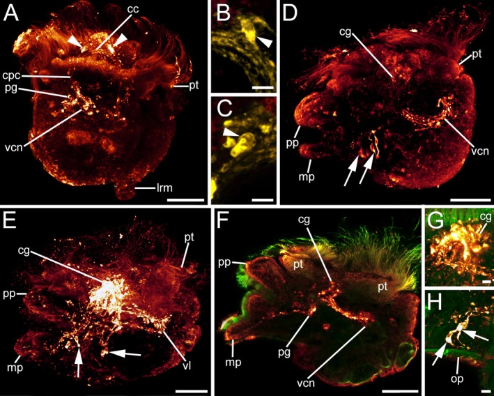
Figure 8.

FMRFamid‐like immunoreactivity (lir) in Lottia cf. kogamogai larvae. FMRFamide‐lir, red A, D–H or yellow B, C; cilia, green. Anterior faces upwards in all aspects. A–C dorsal view; D–H lateral view, ventral to the left. Age of larvae is given in days postfertilization (dpf). A late pediveliger larva (2 dpf), cerebral commissure (cc) between the future cerebral ganglia and with first labelled cells (arrowheads) in each ganglion visible. cpc, cerebropedal connective; lrm, main larval retractor muscle; pg, pedal ganglion; pt, prototroch; vcn, visceral neurite. B same larva as in A magnification of the cell in the right cerebral ganglion anlage. C same larva as in A magnification of the cell in the left cerebral ganglion anlage. D same stage larva as in A. Note the paired flask‐shaped cells (arrows) on each side at the base of the foot and the numerous neurites (vcn) that run from the cerebral ganglia (cg) posteriorly towards the visceral mass. mp, metapodium; pp, propodium. E metamorphic competent larva (3 dpf) with an elaborate nervous system labelled with antibodies to FMRFamide that is composed of strongly stained cerebral ganglia, from which neurites run posteriorly towards the visceral mass forming the visceral loop (vl). F same stage larva as in E optical section showing part of a cerebral and pedal ganglion (pg) from which neurites run towards the visceral mass and into the foot. G same larva as in F magnification of the relatively large future adult, horseshoe‐shaped cerebral ganglia. H same larva as in F magnification of one pair of flask‐shaped cells at the base of the foot. op, operculum. A–H CLSM, maximum projections, except F, which is an optical section. Scale bars: A, D, E, F = 30 μm; B, C, G–H = 5 μm
