Figure 11.

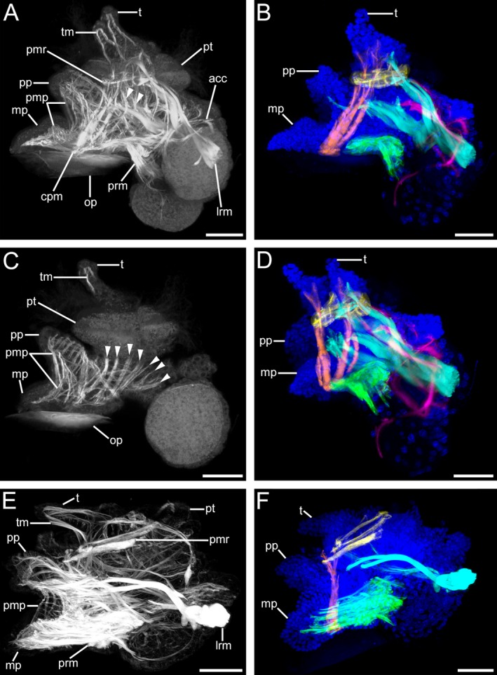
Musculature of Lottia cf. kogamogai larvae. (A, C, E) musculature in white; (B, D, F) muscles colour coded: blue, cell nuclei; cyan, main larval retractor muscle; green, pedal retractor muscle; orange, cephalopedal muscle; pink, accessory retractor muscle; yellow, prototroch muscle ring. (B, D, F) the tentacle, foot and dorso‐ventral musculature are omitted for clarity. Anterior faces upwards in all images. (A‐F) lateral view, ventral to the left. Age of larvae is given in days postfertilization (dpf). (A) late pediveliger larva (2 dpf) showing a prominent main larval retractor muscle (lrm) that runs from the insertion site anteriorly into the prototroch (pt) and the propodium (pp), while branches of the accessory retractor muscles (acc) run along the mantle and to the prototroch. The foot and the tentacles (t) comprise a meshwork of longitudinal and transversal muscle fibres (pmp, tm, respectively). Note the dorso‐ventral muscle fibres (arrowheads) that run from the head and visceral mass towards the foot. cpm, cephalopedal musculature; mp, metapodium; op, operculum; pmr, prototroch muscle ring; prm, pedal retractor muscle. (B) same larva as in (A) showing main muscle systems. Note that the paired cephalopedal muscle inserts at the base of the metapodium and runs towards the base of the respective ipsilateral tentacle. (C) same specimen as in (A), optical section showing the muscular meshwork within the larval foot and numerous dorso‐ventral muscles. (D) same specimen as in (A), slightly tilted to the left, the paired nature of the cephalopedal musculature is visible, which runs through the head, crossing the prototroch muscle ring on the inner side, to each ipsilateral base of the tentacle. Note that the ventral branch of the main larval retractor muscle runs into the more anterior part of the foot, the propodium. (E) metamorphic competent larva (3 dpf) with a dominant pedal retractor muscle, while the accessory retractor muscle is no longer visible and the prototroch muscle ring as well as the main larval retractor muscle are smaller than in the stage before. (F) same specimen as in (E) the prototroch muscle ring and the main larval retractor muscle show signs of disintegration. (A‐F) CLSM, maximum projections, except (C), which is an optical section. Scale bars: (A‐F) = 30 μm
