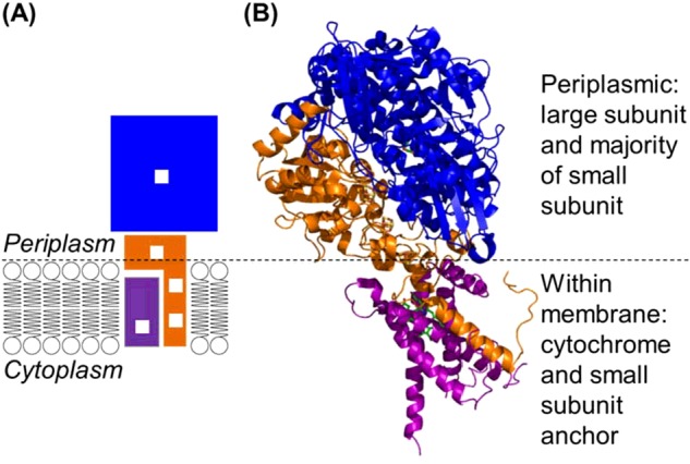
Figure 2. The orientation of a NiFe MBH within a bacterial cell.

(A) Cartoon depiction of how a NiFe MBH is located within the cytoplasmic membrane, with white boxes representing the redox active metal centres and blue, orange and purple blocks indicating the large, small and cytochrome subunits, respectively. (B) Crystallographic insight into how the E. coli hydrogenase-1 large (blue ribbon), small (orange ribbon) and cytochrome (purple ribbon) subunits can interact. Figure generated from PDB 4GD3 [68].
