
Figure 2. Representative images of colorectal cancers stained for VE1.
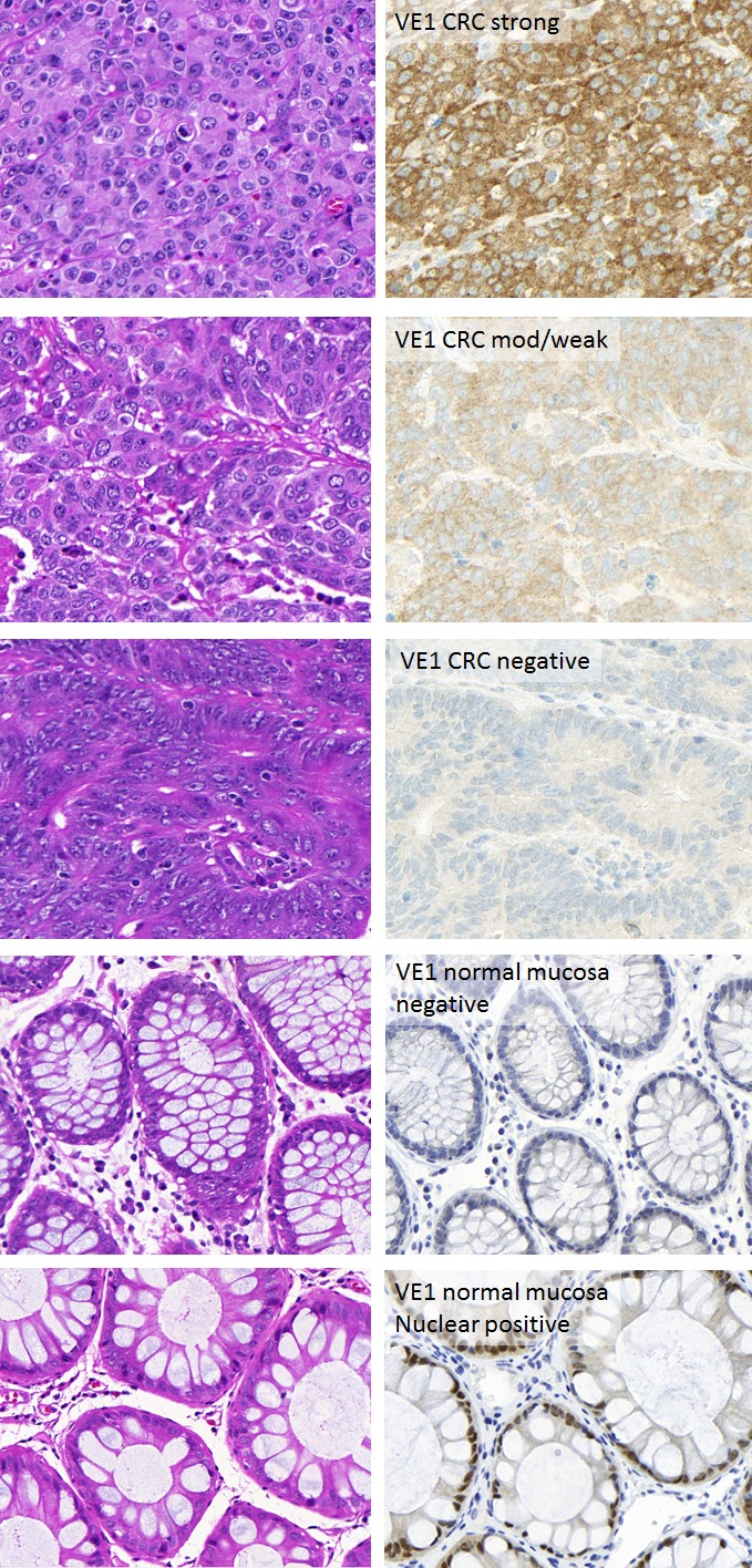
Left panels: Hematoxylin and Eosin of corresponding cases. Right panels: VE1 staining in colorectal cancers and normal mucosa. Staining was in the epithelium and cytoplasmic. Expression ranged from strong to moderate/weak and negative. Normal mucosa was negative for cytoplasmic staining although some nuclear positivity was frequently seen.