
Figure 1. BV6 synergizes with DOX and vinca alkaloids to induce apoptosis in NB cells.
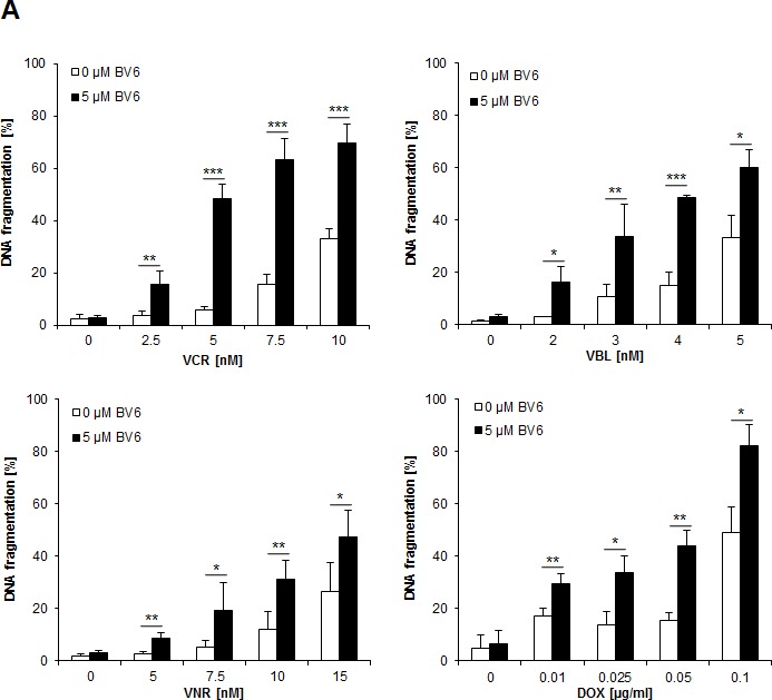

A. SH-EP cells were treated with indicated concentrations of DOX, VCR, VBL, VNR and/or 5 μM BV6 for 72 hours. Apoptosis was determined by analysis of DNA fragmentation of PI-stained nuclei using flow cytometry. Data are shown as mean and SD of three independent experiments performed in triplicate; *, P < 0.05; **, P < 0.01; ***, P < 0.001. B. SH-EP cells were treated with either 5 μM BV6 and/or 0.05 μg/ml DOX or 5 μM BV6 and/or 5 nM VCR for 48 hours. Cell viability was measured by crystal violet assay and is expressed as a percentage of untreated cells. Data are shown as mean and SD of three independent experiments performed in triplicate; *, P < 0.05; **, P < 0.01; ***, P < 0.001. C. SH-EP cells were treated with either 5 μM BV6 for 11 hours and/or 0.05 μg/ml DOX for 1 hour or 5 μM BV6 and/or 5 nM VCR for 72 hours. Colony formation was assessed as described in Material and Methods. The number of colonies is expressed as percentage of controls (upper panels) and representative images are shown (lower panels). Data are shown as mean and SD of three independent experiments performed in triplicate; *, P < 0.05; **, P < 0.01.