
Figure 5. VCR/BV6 cotreatment engages the mitochondrial pathway.
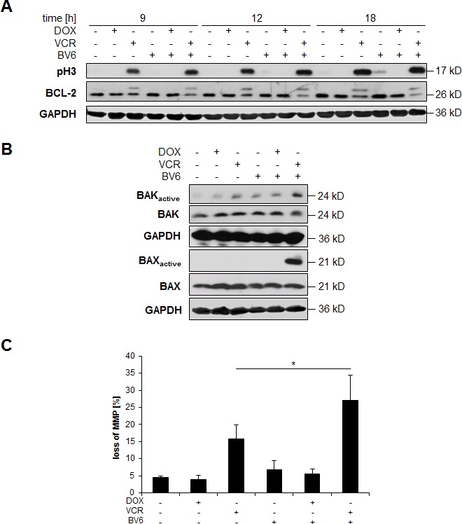
A. SH-EP cells were treated with 5 μM BV6 and/or 0.05 μg/ml DOX and/or 5 nM VCR for indicated times and expression of BCL-2 or phosphorylated histone H3 (pH3) was analyzed by Western blotting; expression of GAPDH served as loading control. B. SH-EP cells were treated with 5 μM BV6 and/or 0.05 μg/ml DOX and/or 5 nM VCR for 18 hours. BAK or BAX were immunoprecipitated using active conformation-specific antibodies and expression of active and total BAK or BAX was analyzed by Western blotting, GAPDH served as loading control. C. SH-EP cells were treated with 5 μM BV6 and/or 0.05 μg/ml DOX and/or 5 nM VCR for 18 hours. MMP was analyzed by flow cytometry using JC-1 staining. Data are shown as mean and SD of three independent experiments performed in triplicate; *, P < 0.05.