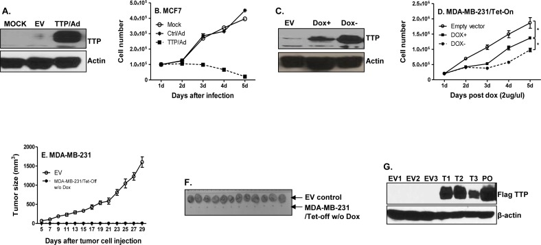
Figure 1. TTP inhibits breast cancer cell proliferation and tumor development.
MCF7 cells were infected with TTP/adenovirus and control adenovirus at MOI=1. TTP expression was detected 24 h after infection by Western blot with anti-TTP antibody A., and cell numbers were counted every 24 h until 5 days after infection B. Results shown are mean plus SEM of three independent experiments with each run in duplicate. 1 × 10⁵ TTP/Tet-Off MDA-MB-231 cells were cultured with or without 2 μg/ml doxycycline (Dox). TTP expression was measured by western blot 5 days after withdraw Dox C., and cell counting was performed at indicated times D. TTP/Tet-Off MDA-MB-231 cells were cultured for one week without Dox, and then 5 × 10⁶ TTP/Tet-Off MDA-MB-231 were inoculated s.c. into mammary glands of the NSG mice. Tumor growth was measured and recorded E. Tumors were excised at day 29 after tumor cell inoculation and representative tumors for each experimental group were shown F., G. Tumor tissues were lysed and total proteins were extracted for detecting Flag-tagged TTP levels by western blot with anti-FLAG antibody. EV: tumors induced with Tet-off cells expressing empty vector; T: tumors generated with Tet-off cells expressing TTP. Number means the number of tumors.

