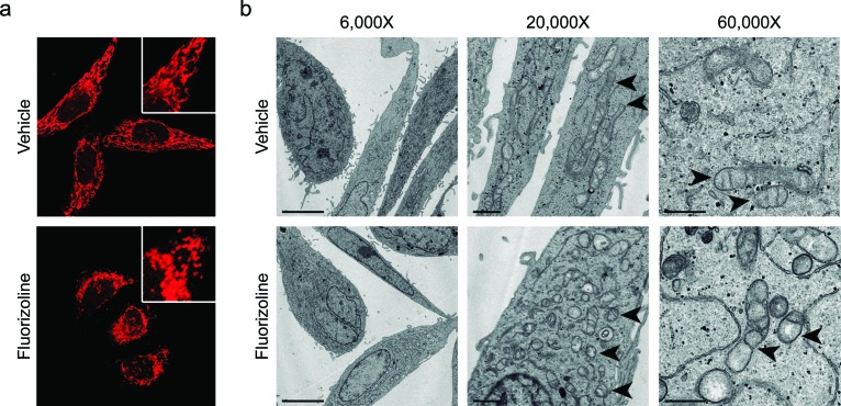
Figure 2. Treatment with fluorizoline induces changes in the mitochondrial morphology and ultrastructure.
a. HeLa cells were treated with DMSO (vehicle) or 10 μM fluorizoline for 4 h. Mitochondria were stained with 100 nM MitoTracker® Red CMXRos and imaged using a confocal microscope. b. HeLa cells were incubated with DMSO (vehicle) or 2 μM fluorizoline for 24 h and changes in the mitochondrial morphology were visualized by transmission electron microscopy. Magnification at 6,000X (scale corresponding to 5 μm), 20,000X (scale corresponding to 1 μm), and 60,000X (scale corresponding to 0.5 μm). Arrowheads indicate the mitochondria.

