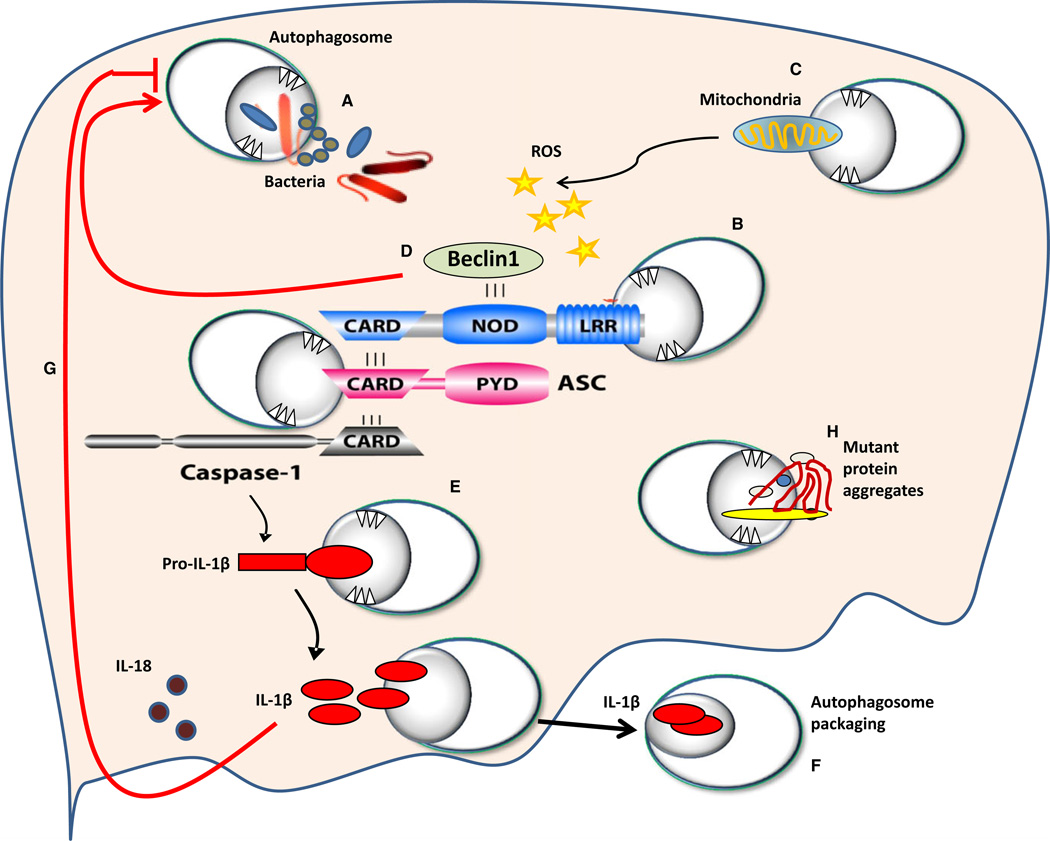
Fig. 1. Schematic representation of molecular interaction between autophagosomes and members of the inflammasome.
NOD-like receptors (NLRs) sense bacterial or viral molecules leading to the assembly of the inflammasome complex. (A) Autophagy can uptake certain invading organisms. The inflammasome complex is composed of the NLR, the adapter molecule ASC, and caspase-1 (B). Members of the inflammasome are ubiquitinated and then targeted by autophagosomes (B). The inflammasome can be activated by reactive oxygen species (ROS) released from mitochondria that were not cleared by autophagosomes (C). NLRs bind beclin-1, and upon their recruitment to the inflammasome complex, beclin-1 is released and contributes to the formation of autophagosomes (D). Autophagosomes also target pro-IL-1β to maintain its level under control (E). The inflammasome complex cleaves pro-IL-1β leading to active IL-1β. Active IL-1β is escorted outside the cell within autophagosomes (F). IL-1β inhibits autophagosome formation via unknown mechanisms (G). Mutant protein aggregates are targeted by autophagy to clear them from the cytosol; however, if they accumulate, autophagy molecules will be sequestered reducing their availability to form autophagosomes (H). Red arrows depict pathways by which the inflammasome contributes to the modulation of autophagy.

