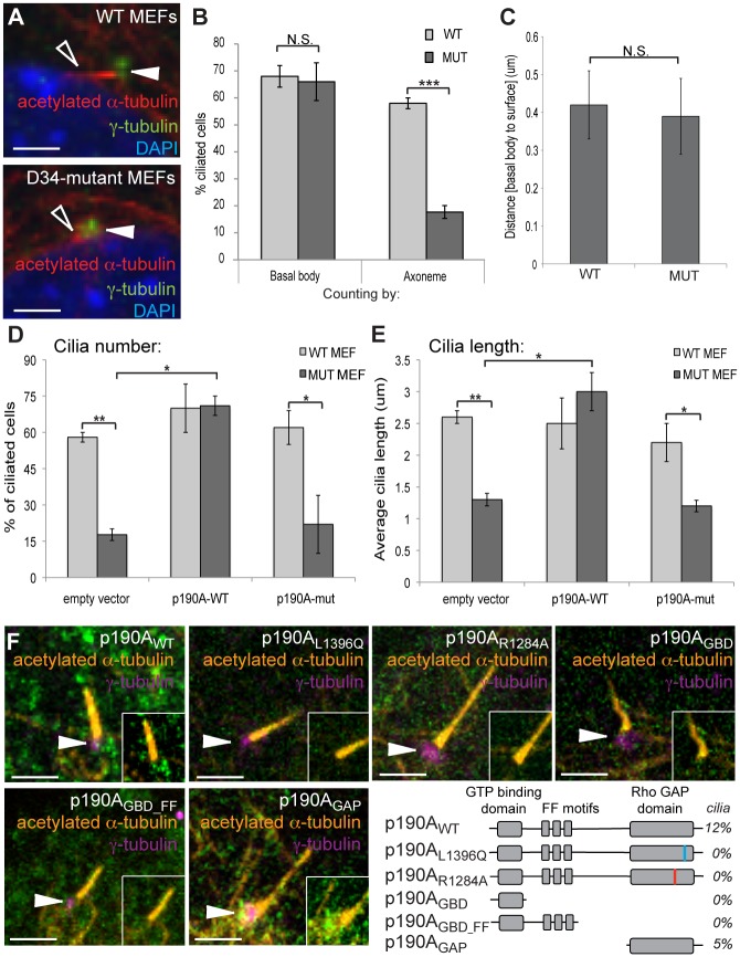
Fig 5. Mouse embryonic fibroblasts (MEFs) derived from Arhgap35D34/D34 embryos exhibit defects in cilia elongation, associated with a failure of p190AL1396Q to be recruited to the base of the cilium.
(A) Immunofluorescent staining of MEFs stimulated to form cilia by serum withdrawal reveals a defect in cilia length (acetylated α-tubulin, open arrowhead) independent of the basal body (γ-tubulin, closed arrowhead). (B) Quantification of number of ciliated cells based on basal body (γ-tubulin) and axoneme (acetylated α-tubulin). (C) The positioning of the basal body with respect to the cell surface is similar between control and Arhgap35D34/D34 MEFs. (D-E) The defects in cilia number (panel D) and length (panel E) in Arhgap35D34/D34 MEFs can be rescued by introduction of full-length wild-type p190A, but not the p190AL1396Q mutant protein. (F) Full-length GFP-tagged p190AWT and p190AGAP constructs are enriched at the basal body (marked by γ-tubulin; arrowheads) while p190AL1396Q, P190AR1284A, p190AGBD, and p190AGBD_FF constructs fail to specifically localize in wild type MEFs. The percentage of cells with ciliary localization is indicated (N>200 cells per construct), which suggests a transient recruitment to the basal body. Scale bars, 2.5μm. *p<0.05, **p<0.01, ***p<0.005 (one-way ANOVA)

