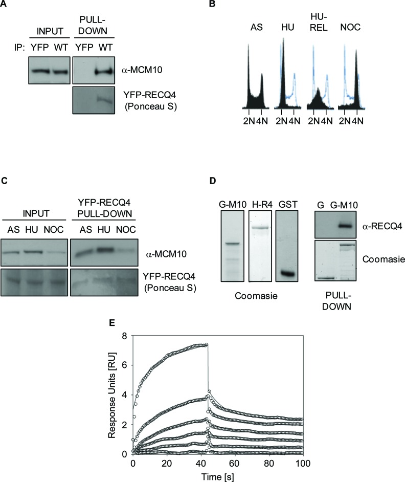
Figure 1. RECQ4 interacts directly with MCM10.
A. Immunoblot analysis for MCM10 in anti-YFP immunoprecipitates from U20S cells expressing either YFP alone or YFP-RECQ4 (WT). Ponceau S staining was used to confirm the presence of YFP-RECQ4 in the precipitates. B. Flow cytometry analysis for asynchronous (AS), hydroxyurea-arrested (HU), HU-arrested cells released into S-phase (HU-REL) and nocodozole arrested U20S cells (NOC). DNA was stained using propidium iodide. The solid black traces show the distribution of cells, and the blue traces show the outline of the control (AS) sample for comparison. C. Immunoblot analysis for MCM10 in anti-YFP-RECQ4 immunoprecipitates from the cells shown in panel B. Loading is shown using Ponceau S staining. D. Coomassie blue stained polyacrylamide gels showing purified GST-MCM10 (G-M10), His-RECQ4 (H-R4) or GST alone (G and GST). The panel on the right shows immunoblot analysis for RECQ4 in GST pull-down samples, together with a Coomassie stained gel showing the loading control for the GST proteins. E. Binding kinetics analysis of RECQ4 interacting with immobilized MCM10 using surface plasmon resonance (Biacore). Open circles represent the experimental data at different RECQ4 concentrations (1, 0.5, 0.25, 0.125, 0.06, and 0.03 μM) and the solid lines represent the fit of the data to a two state model. The dissociation constant (Kd) was determined by the ratio of kon and koff and was found to be 170±200 nM.

