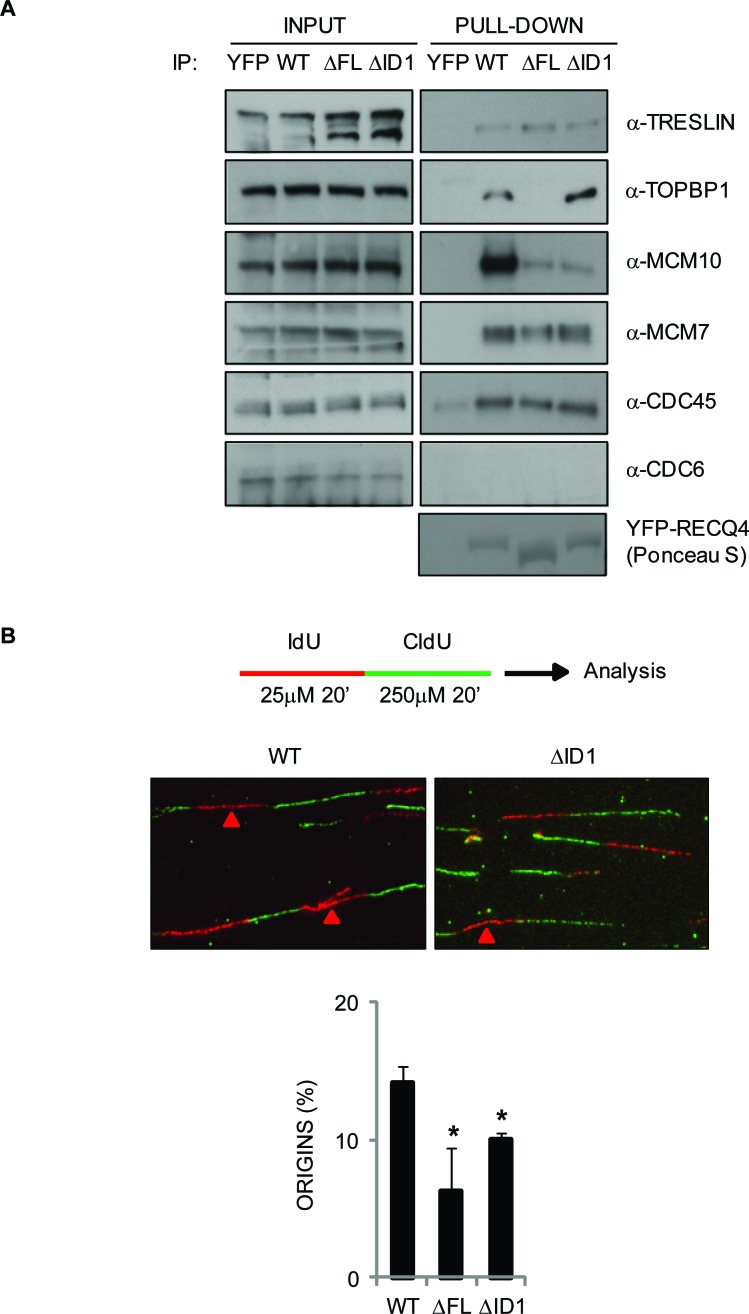
Figure 6. Analysis of protein complexes associated with the truncated RECQ4 variants and their ability to support normal DNA replication in DT40 cells.
A. Western blotting analysis of proteins pulled-down using YFP alone, YFP-RECQ4, or the ΔFL and ΔID1 variants. The input samples for each pull down are shown. The proteins analyzed are shown on the right. Ponceau S staining was used to confirm a similar loading of the YFP-RECQ4 proteins. B. DNA replication analysis on isolated DNA fibres. The upper cartoon shows the sequential labeling protocol using IdU (red) and CldU (green) nucleoside analogues. The middle panel shows representative fibres. Red arrows devote red tracts flanked by green tracts, denoting the positions of replication origins. The lower panel shows relative frequency of origin firing events. Error bars represent SD from three independent experiments. *=p < 0.05.

