
Figure 2. Immunohistochemistry based morphoproteomic studies of osteosarcoma Patient #1.
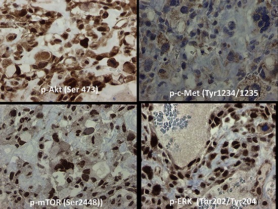
In the tumor cells of Patient #1, c-Met was expressed at 0–1+ (rarely 2+) in the cytoplasm, mTOR at 3+ in nucleus and cytoplasm, ERK 1/2 at 3+ in the nucleus, and Akt at 0–3+ in both nucleus and cytoplasm. Representative sections are shown.