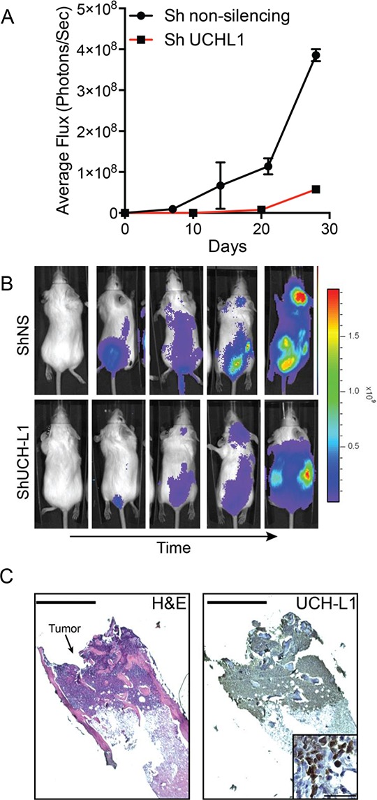
Figure 5. UCH-L1 depletion impairs the dissemination of myeloma in an orthotopic model.

A. Bioluminescence imaging (BLI) of SCID-Beige mice (n = 3 each condition) following i.v. injection of KMS-11LUC cells transduced with CMV driven shRNAs as indicated. The graph represents the mean +/− SEM at each time point. B. Representative images of mice from (A) C. Histologic appearance of a myeloma lesion in the femur of an affected mouse. Paraffin embedded sections were stained with hematoxylin and eosin (H&E) or a UCH-L1 specific antibody followed by development with horseradish peroxidase (HRP). Bar = 400 μm. The inset is a high-power view of the same sample, bar = 50 μm.
