
Figure 2. CUDR overexpression cooperated with cyclinD1 overexpression or PTEN depletion accerlates the liver cancer stem cell proliferation.
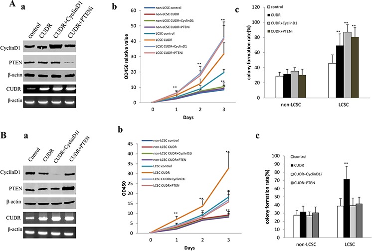
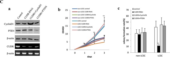
A. The growth and colony formation ability in the stable human liver cancer stem cell(HLCSC) lines and non-HLCSC transfected with pCMV6-A-GFP, pCMV6-A-GFP-CUDR, pCMV6-A-GFP-CUDR plus pcDNA3.1-CyclinD1, pCMV6-A-GFP-CUDR plus pGFP-V-RS-PTEN, respectively. a. RT-PCR analysis of CUDR mRNA and Western bloting with anti-cyclinD1, anti-PTEN expression in stable liver cancer stem cells transfected with pCMV6-A-GFP, pCMV6-A- GFP-CUDR, pCMV6-A-CUDR plus pcDNA3.1-CyclinD1, pCMV6-A- GFP-CUDR plus pGFP-V-RS-PTEN, respectively (indicated in the left). β-actin as internalcontrol. b. Cell proliferation assay in vitro in liver cancer stem cells and unstemic liver cancer. Data are means of value from three independent experiment, bar ± SEM. ** P < 0.01; * P < 0.05. c. Cells colony-formation efficiency assay in liver cancer stem cells and unstemic liver cancer cells. Data are means of value from three independent experiment, bar ± SEM. ** P < 0.01; * P < 0.05. B. The growth and colony formation ability in the stable human liver cancer stem cell (HLCSC) lines and non-HLCSC transfected with pCMV6-A-GFP, pCMV6-A-GFP-CUDR, pCMV6-A-GFP-CUDR plus pGFP-V-RS—CyclinD1, pCMV6-A-GFP-CUDR plus pcDNA3.1-PTEN, respectively. a. RT-PCR analysis of CUDR mRNA and Western bloting with anti-cyclinD1, anti-PTEN expression in stable liver cancer stem cells transfected with pCMV6-A-GFP, pCMV6-A-GFP-CUDR, pCMV6-A- GFP-CUDR plus pGFP-V-RS-CyclinD1, pCMV6-A-GFP-CUDR plus pcDNA3.1-PTEN, respectively (indicated in the left). β-actin as internalcontrol. b. Cell proliferation assay in vitro in liver cancer stem cells and unstemic liver cancer cells. Data are means of value from three independent experiment, bar ± SEM. ** P < 0.01; * P < 0.05. c. Cells colony-formation efficiency assay in liver cancer stem cells and unstemic liver cancer cell. Data are means of value from three independent experiment, bar ± SEM. ** P < 0.01; * P < 0.05. C. The growth and colony formation ability in the stable human liver cancer stem cell(HLCSC) lines and non-HLCSC transfected with pGFP-V-RS, pGFP-V-RS-CUDR, pGFP-V-RS-CUDR plus pcDNA3.1-CyclinD1, pGFP-V-RS-CUDR plus pGFP-V-RS-PTEN, respectively. a. RT-PCR analysis of CUDR mRNA and Western bloting with anti-cyclinD1, anti-PTEN expression in stable liver cancer stem cells transfected with pGFP-V-RS, pGFP-V-RS-CUDR, pGFP-V-RS-CUDR plus pcDNA3.1-CyclinD1, pGFP-V-RS-CUDR plus pGFP-V-RS-PTEN, respectively (indicated in the left). β-actin as internalcontrol. b. Cell proliferation assay in vitro in liver cancer stem cells and unstemic liver cancer cells. Data are means of value from three independent experiment, bar ± SEM. **, P < 0.01; *, P < 0.05. c. Cells colony-formation efficiency assay in liver cancer stem cells and liver cancer unstemic cells. Data are means of value from three independent experiment, bar ± SEM. ** P < 0.01; * P < 0.05.