
Figure 2. Fatostatin inhibits cell proliferation and colony formation in PCa cells harboring different p53 status.
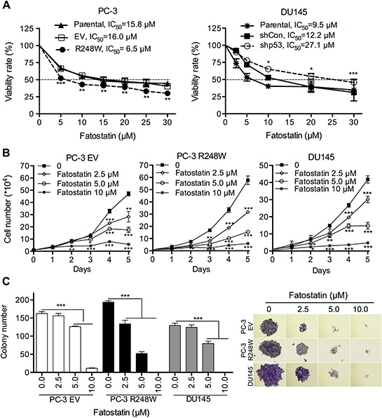
A. Fatostatin suppressed the proliferation of PCa cells of different p53 status. Cells (PC-3 Parental, EV, R248W; DU145 Parental, shCon, shp53) were treated with various concentrations of fatostain for 72 hours and the IC50 values were calculated and shown from three independent experiments. *P < 0.05, **P < 0.01 and ***P < 0.001. B. Fatostatin inhibited growth of PC-3 EV, PC-3 R248W and DU145 cells in a time- and dose-dependent manner. Cell growth was determined by counting cell numbers daily using a hemocytometer. *P < 0.05, **P < 0.01 and ***P < 0.001. C. Fatostatin decreased colony formation of PC-3 EV, PC-3 R248W and DU145 cells in a dose-dependent manner after 10-day treatment. The number of colonies was counted and reported as the mean ± SD of triplicate experiments. ***P < 0.001. Colony development images are shown in the right panel.