
Figure 3. Fatostatin causes G2/M cell cycle arrest and induces apoptosis in PCa cells harboring p53 mutations.
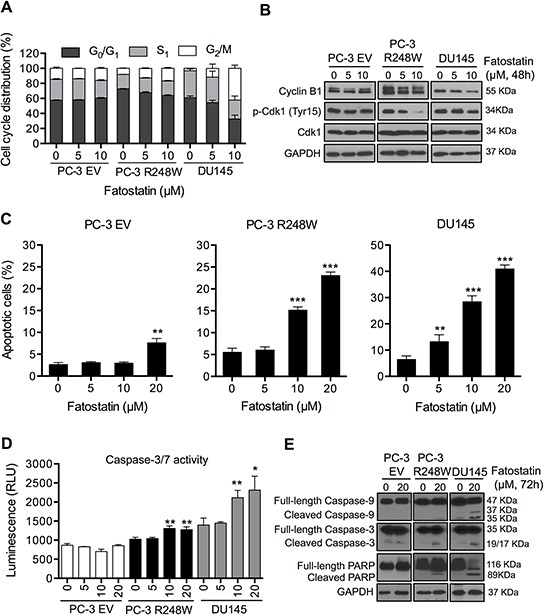
A. Fatostatin caused G2/M cell cycle arrest after 48-hour treatment compared with vehicle in PCa cells harboring p53 mutations. Cell cycle distributions of PC-3 EV, PC-3 R248W and DU145 cells treated with fatostatin or vehicle were assessed by flow cytometry. Data represent the means ± SD values from triplicate experiments. B. Fatostatin decreased the expression of Cyclin B1 and p-Cdk1 (Tyr15) in PC-3 R248W and DU145 cells but not in PC-3 EV cells as determined by Western blot. C. Apoptosis in PC-3 EV, PC-3 R248W and DU145 cells treated by fatostatin was determined by flow cytometry-based Annexin V-FITC and PI staining analysis. **P < 0.01, ***P < 0.001. D. Caspase-3/7 activity in PC-3 EV, PC-3 R248W and DU145 cells treated with vehicle or fatostatin for 48 hours was determined by an enzymatic activity assay. Data are plotted as the relative units of luciferase intensity and reported as the means ± SD values from triplicate experiments. *P < 0.05, **P < 0.01. E. Western blot analysis of apoptosis-related markers (caspase-9, caspase-3 and PARP) in PC-3 EV, PC-3 R248W and DU145 cells treated with 20 μM of fatostatin for 72 hours. GAPDH was used as a loading control.