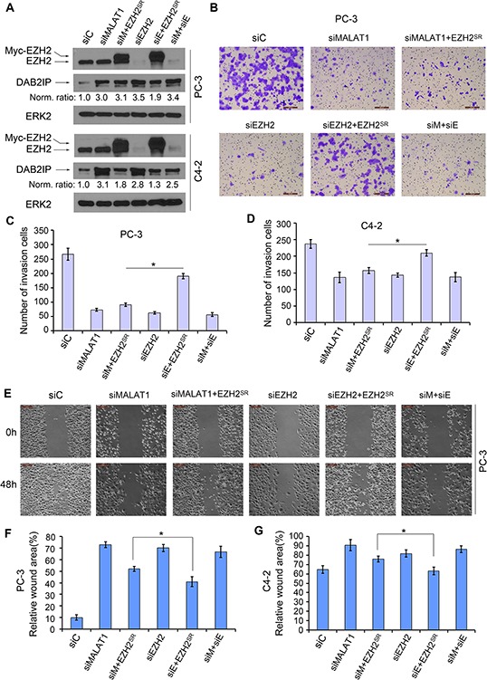
Figure 5. MALAT1 facilitates EZH2-mediated PCa cell migration and invasion.

A. PC-3 and C4-2 cells were transfected with non-specific control (siC), MALAT1- and/or EZH2-specific siRNAs (siM and siE, respectively) in combination with siRNA-resistant EZH2 expression vector (EZH2SR). At 48 h after transfection, cells were harvested for western blot analysis using the indicated antibodies. ERK2 was used as a loading control. The western blot density of DAB2IP was first normalized to the density of ERK2 in each lane and then normalized further to the value in cells transfected with siC. B–D. Cells were transfected as in (A). At 48 h after transfection, cells were used for Matrigel invasion assays. Representative images of invasion assay performed in PC-3 cells are shown in (B) and the quantification results from PC-3 and C4-2 cells are shown in (C) and (D), respectively. Scale bar, 200 μm. Data are means ± S.D. from experiments with three replicates. *P < 0.01. E–G. PC-3 and C4-2 cells were transfected with indicated siRNAs and plasmids as in (A). At 24 h after transfection, artificial wounds were created on cells grown in confluence. Images were taken at 0, 48 h after wound. Representative images from PC-3 cells are shown in (E) and the quantification results are shown in (F) and (G). Scale bar, 50 μm. Data are shown as means ± S.D. from three individual experiments. *P < 0.01.
