
Figure 2. Detection of UbcH8 expression in NPC biopsies by immunohistochemistry.
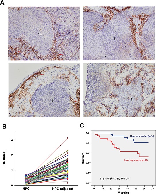
A. Representative slides of NPC samples stained by anti-UbcH8 antibody. Black arrows denote NPC tissue and red arrows denote adjacent stromal tissue. IHC indexes of NPC vs. NPC adjacent: upper left (0.39 vs. 1.18); upper right (0.18 vs. 0.51); lower left (0.53 vs. 1.75); lower right (0.57 vs. 1.40). B. IHC indexes of all the investigated NPC biopsies with paired cancer nest and corresponding adjacent non-cancerous stromal tissue (n = 69). C. Kaplan-Meier survival curves illustrating the significance of UbcH8 expression in NPC. NPC patients with low UbcH8 expression had shorter NPC-specific survival than those with high UbcH8 expression (p = 0.011).