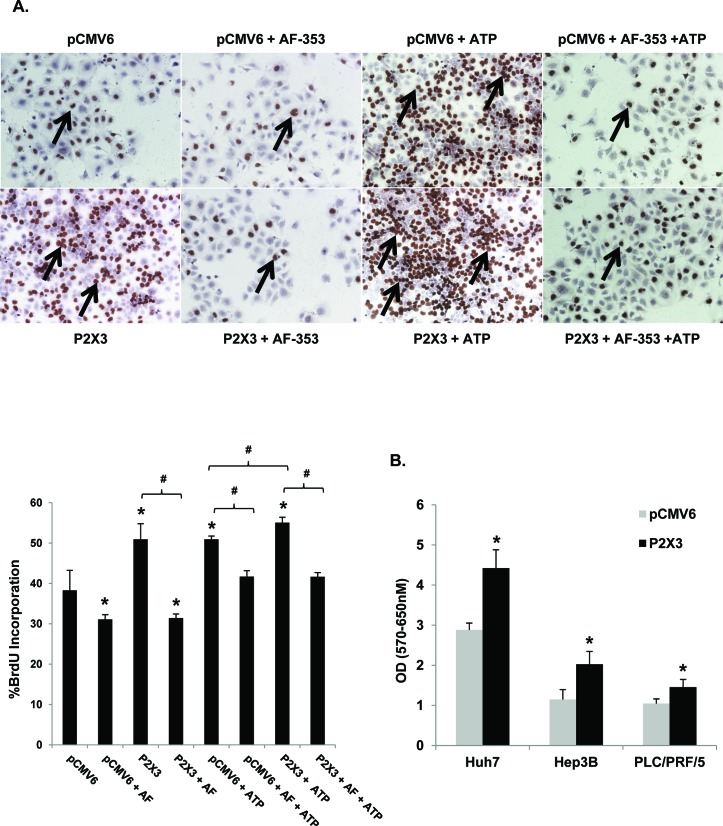
Figure 6. P2X3 overexpression induces basal and ATP mediated proliferation.
A. Light microscopic images (10X) of BrdU immunostained Huh7 cells after pCMV6 plasmid or P2X3 DNA transfection ± ATP (100 μM, 24 hr) ± pre-treatment with AF- 353 (5 μM). Arrows point to BrdU-positive cells, expressed as a percentage of total cells. B. HCC cell proliferation after 72h pCMV6 plasmid or P2X3 DNA transfection, assessed by MTT assay. Data represents mean ± SEM, n = 3-6, *p < 0.05 vs. untreated.

