
Figure 5. Representative results of immunostaining for β-catenin, Notum, Glypican-1, Glypican-3 in FFPE mouse tissue sections.
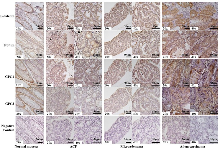
Membrane-bound β-catenin, Notum and Glypican-1 were observed in normal colon mucosa, whereas dysplastic ACF (Aberrant Crypt Foci), microadenomas, and adenocarcinomas exhibited more intense (++) staining (nuclear or cytoplasmic). Glypican-3 staining was less intense (+) than Glypican-1 staining (++), and the adenocarcinoma showed a negative signal (−) with respect to nuclear or cytoplasmic compartments along with more intense (++) extracellular staining. Incubation with a primary antibody was omitted in the negative control. Sections were counterstained with hematoxylin: 20x and 40x original magnification. Scale bar, 50 μm.