
Figure 2. SPC24 protein was analyzed by immunohistochemistry and western blot in HCC tissues.
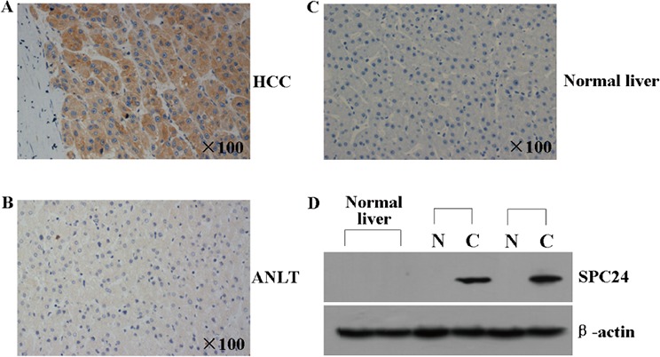
A and B. The representative immunohistochemical pictures of one pair of HCC specimen (upper panel) and corresponding noncancerous tissue (lower panel) from a tissue array containing 69 pairs of HCC specimens were shown. The pictures were stainied with SPC24 antibody, and the nuclei were counterstained with hematoxylin. C. The representative immunohistochemical staining in one normal liver tissue is shown. The nuclei were counterstained with hematoxylin. D. SPC24 expression in 2 normal liver tissues, 2 paired HCC [C] and adjacent non-cancerous liver tissues [N] was evaluated by Western blot. β-actin protein was used as an internal control.