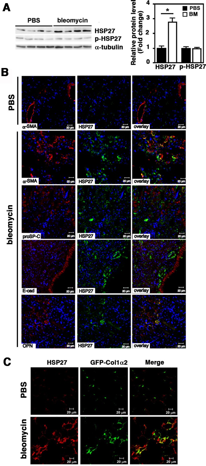
Fig 3. Upregulation of HSP27 in lung tissues of bleomycin-treated mice.

Mice were intratracheally treated with PBS or bleomycin. After 14 days, mice were sacrificed and lungs were removed. (A) Immunoblot analysis. Protein levels of HSP27 and p-HSP27 were analyzed by Immunoblotting using tissue lysates prepared from right lungs. For a loading control, α-tubulin was used. Signal intensities were quantified using Image J software. A representative image from seven independent experiments is shown in the left. Quantitative data are shown as mean ± SE (n = 7) in the right. *: P<0.05 by Student’s t-test. (B) Immunofluorescence staining. Left lungs were fixed with 10% formaldehyde and embedded in paraffin. Tissue sections (4 μm) were double stained for HSP27 (green) and α-SMA (red), proSP-C (red), E-cadherin (E-cad, red) or OPN (red) as depicted. For nuclear staining, TO-PRO-3 (blue) was used. The bars indicate 20 μm. Representative images from three independent experiments are shown. (C) Col1a2-EGFP reporter mice. Mice were intratracheally instilled with PBS or bleomycin. After 14 days, mice were sacrificed and lungs were fixed with 4% paraformaldehyde, treated with 30% sucrose for cryoprotection, and embedded. Frozen sections (6 μm thick) were stained for HSP27 (red). Collagen Type I α2 was visualized by EGFP (green). The bars indicate 20 μm. Representative images from three independent experiments are shown.
