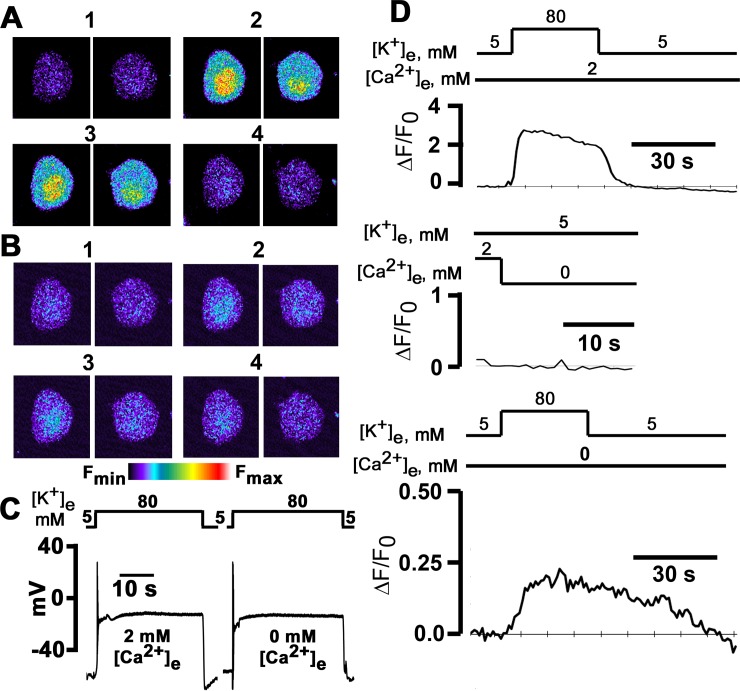
Fig 1. High [K+]e-induced intracellular Ca2+ transients in the presence and absence of extracellular Ca2+.
A and B: x-y fluorescence images recorded in 24 h cultured sympathetic ganglion neurons loaded with fluo-4/AM before, during and following 30-s depolarizations with 80 mM [K+]e in 2 mM [Ca2+]e (A) and in the absence of extracellular Ca2+ with 200 μM EGTA added to the extracellular solution (B). Exposure to elevated [K+]e evoked transient increases in both cyto- and nucleoplasmic Ca2+ concentrations. Images in panels 2 and 3 were acquired at 10 and 30 s after initiating the [K+]e challenge. C: High [K+]e depolarizes the membrane potential both in the presence and absence of external Ca2+: 80 mM [K+]e rapidly depolarized Vm from a resting membrane potential of -44 mV to 1 mV in 2 mM [Ca2+]e, and from -41 mV to 0 mV in the absence of extracellular Ca2+. The level of depolarization was sustained for the duration of the [K+]e challenge, both in the presence and absence of external Ca2+. D: Time courses of fractional fluorescence (ΔF/F0), i.e., [Ca2+]i, in the cytoplasm during exposure to 80 mM [K+]e in the presence and absence of external Ca2+. ΔF/F0 was calculated from the fluorescence intensity measured in the whole cytoplasm for each image taken every second.

