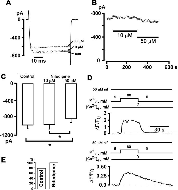
Fig 6. The 1,4-dihydropyridine antagonist of L-type Ca2+ channels, nifedipine, does not affect Ca2+ transients elicited by exposure to elevated [K+]e.
A and B: The response of peak IBa in a postganglionic sympathetic neuron to application of 10 and 50 μM nifedipine. IBa was activated by depolarizations from -80 mV to 10 mV every 10 seconds. Representative current traces are shown in A. C: Bar graphs represent the average peak IBa in control and in 10 and 50 μM nifedipine. Values are from 10 cells. *P < 0.05, RM ANOVA on ranks followed by Student-Newman-Keuls Method for multiple comparisons. D: Exemplar time courses of changes in cytosolic ΔF/F0 elicited by 80 mM [K+]e in the presence (upper panel) and absence of external Ca2+. The cell was continuously bathed in 50 μM nifedipine starting 20 min before the first [K+]e test. E: Percentage of cells exhibiting [Ca2+]i transients both in 2 mM and 0 mM [Ca2+]e; P = non-significant versus control by Fisher Exact test (60 cells for control and 10 cells for nifedipine).

