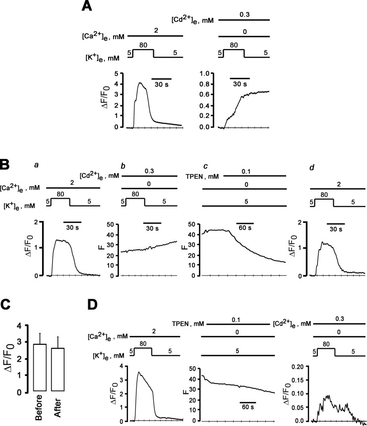
Fig 7. External Cd2+ does not suppress high [K+]e-induced ΔF/Fo transients in 0 mM [Ca2+]e.
A: Irreversible increase of ΔF/F0 in depolarized neurons in the presence of extracellular Cd2+. Shown are time courses of changes in cytosolic ΔF/F0 elicited by 80 mM [K+]e in the presence of 2 mM Ca2+ (left panel) and by 80 mM [K+]e and 300 μM Cd2+ in a Ca2+-free bath solution (right panel). The increase of ΔF/F0 does not recover after removal of high-[K+]e-induced depolarization. B: External cadmium does not block depolarization-induced increase in cytosolic ΔF/F0 in the absence of extracellular Ca2+. (a) reversible increase in ΔF/F0 in response to a 30-s exposure to 80 mM [K+]e in 2-mM Ca2+ bath solution. (b) and (c): a second exposure to 80 mM [K+]e in Ca2+-free solution supplemented with 300 μM CdCl2 causes a sustained increase in fluo-4 fluorescence (b) which is not reversed until treatment of the cell with the membrane permeable metal chelator TPEN at 100 μM (c). A second K+ challenge of the TPEN-loaded neuron in a 2-mM Ca2+ bath solution (d) shows restoration of fluo-4 Ca2+ responsivity. C: Bar graphs summarizing mean ± SEM of peak cytosolic ΔF/F0 in sympathetic neurons before and after loading with TPEN (100 μM). P = 0.14 by paired t-test (n = 5 cells). D: Extracellular cadmium does not block depolarization-induced increase in ΔF/F0 in the absence of external Ca2+. Left panel: reversible increase in ΔF/F0 in response to a 30-s exposure to 80 mM [K+]e in 2 mM [Ca2+]e. Following TPEN loading (middle panel), a second 30-s [K+]e challenge in a Ca2+-free bath solution containing 300 μM Cd2+ gives rise to a small-amplitude ΔF/F0 transient (right panel).

