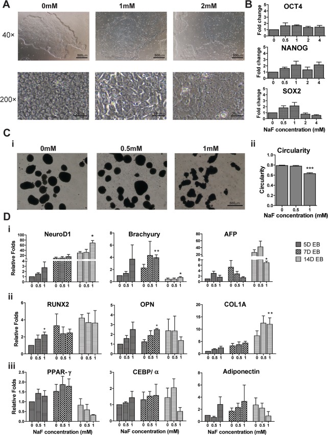
Fig 1. Sodium Fluorides (NaF) affected the differentiation of H9 hESCs.
(A) The morphology of hESCs was characterized under an inverted microscope. NaF-treated hESCs were larger and flattened than untreated cells were. (B) The expressions of pluripotent markers (OCT4, NANOG and SOX2) in untreated and NaF-treated hESCs were comparable, as quantified by real-time polymerase chain reaction (PCR). (C) The morphology of embryoid bodies (EBs) derived from hESCs after 5 days (5D) of differentiation in the presence or absence of NaF. (i) 5D EBs in untreated and 0.5 mM NaF-treated groups exhibited a circular in shape while EBs in the 1 mM NaF-treated group were irregular in shape. (ii) The circularities of 5D EBs in the 1 mM NaF group were significantly lower than in the untreated and 0.5 mM NaF-treated groups. (D) The gene expression patterns of EBs were disturbed by high-dose NaF treatment. (i) The expression of three germ layer markers (Ectoderm: NeuroD1, Mesoderm: Brachyury, Endoderm: AFP) in EBs. (ii) Expression of osteogenesis markers (RUNX2, OPN and COL1A) in EBs. (iii) Expression of adipogenesis markers (PPAR-γ, CEBP/α, Adiponectin) in EBs. 40×: 40× magnification. 200×: 200× magnification. *, p < 0.05. **, p < 0.01. ***, p < 0.001.

